Zinc »
PDB 1ylk-1z5h »
1ym3 »
Zinc in PDB 1ym3: Crystal Structure of Carbonic Anhydrase RV3588C From Mycobacterium Tuberculosis
Enzymatic activity of Crystal Structure of Carbonic Anhydrase RV3588C From Mycobacterium Tuberculosis
All present enzymatic activity of Crystal Structure of Carbonic Anhydrase RV3588C From Mycobacterium Tuberculosis:
4.2.1.1;
4.2.1.1;
Protein crystallography data
The structure of Crystal Structure of Carbonic Anhydrase RV3588C From Mycobacterium Tuberculosis, PDB code: 1ym3
was solved by
A.S.Covarrubias,
A.M.Larsson,
M.Hogbom,
J.Lindberg,
T.Bergfors,
C.Bjorkelid,
S.L.Mowbray,
T.Unge,
T.A.Jones,
Structural Proteomics Ineurope (Spine),
with X-Ray Crystallography technique. A brief refinement statistics is given in the table below:
| Resolution Low / High (Å) | 26.20 / 1.75 |
| Space group | P 41 21 2 |
| Cell size a, b, c (Å), α, β, γ (°) | 56.377, 56.377, 104.192, 90.00, 90.00, 90.00 |
| R / Rfree (%) | 17.9 / 22.9 |
Other elements in 1ym3:
The structure of Crystal Structure of Carbonic Anhydrase RV3588C From Mycobacterium Tuberculosis also contains other interesting chemical elements:
| Magnesium | (Mg) | 1 atom |
Zinc Binding Sites:
The binding sites of Zinc atom in the Crystal Structure of Carbonic Anhydrase RV3588C From Mycobacterium Tuberculosis
(pdb code 1ym3). This binding sites where shown within
5.0 Angstroms radius around Zinc atom.
In total only one binding site of Zinc was determined in the Crystal Structure of Carbonic Anhydrase RV3588C From Mycobacterium Tuberculosis, PDB code: 1ym3:
In total only one binding site of Zinc was determined in the Crystal Structure of Carbonic Anhydrase RV3588C From Mycobacterium Tuberculosis, PDB code: 1ym3:
Zinc binding site 1 out of 1 in 1ym3
Go back to
Zinc binding site 1 out
of 1 in the Crystal Structure of Carbonic Anhydrase RV3588C From Mycobacterium Tuberculosis
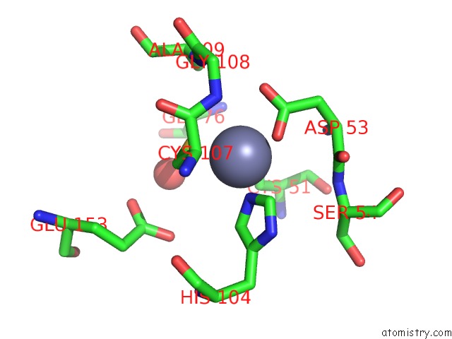
Mono view
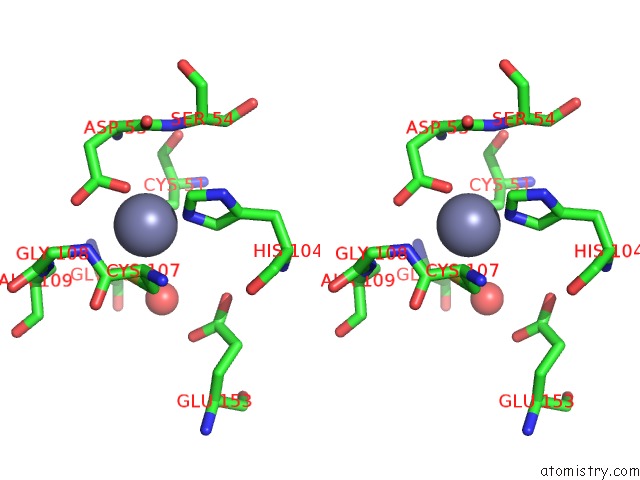
Stereo pair view
Mono view
Stereo pair view
A full contact list of Zinc with other atoms in the Zn binding
site number 1 of Crystal Structure of Carbonic Anhydrase RV3588C From Mycobacterium Tuberculosis within 5.0Å range:
|
Reference:
A.Suarez Covarrubias,
A.M.Larsson,
M.Hogbom,
J.Lindberg,
T.Bergfors,
C.Bjorkelid,
S.L.Mowbray,
T.Unge,
T.A.Jones.
Structure and Function of Carbonic Anhydrases From Mycobacterium Tuberculosis. J.Biol.Chem. V. 280 18782 2005.
ISSN: ISSN 0021-9258
PubMed: 15753099
DOI: 10.1074/JBC.M414348200
Page generated: Wed Oct 16 20:59:45 2024
ISSN: ISSN 0021-9258
PubMed: 15753099
DOI: 10.1074/JBC.M414348200
Last articles
Zn in 9MJ5Zn in 9HNW
Zn in 9G0L
Zn in 9FNE
Zn in 9DZN
Zn in 9E0I
Zn in 9D32
Zn in 9DAK
Zn in 8ZXC
Zn in 8ZUF