Zinc »
PDB 1y8q-1ykf »
1yhu »
Zinc in PDB 1yhu: Crystal Structure of Riftia Pachyptila C1 Hemoglobin Reveals Novel Assembly of 24 Subunits.
Protein crystallography data
The structure of Crystal Structure of Riftia Pachyptila C1 Hemoglobin Reveals Novel Assembly of 24 Subunits., PDB code: 1yhu
was solved by
J.F.Flores,
C.R.Fisher,
S.L.Carney,
B.N.Green,
J.K.Freytag,
S.W.Schaeffer,
W.E.Royer,
with X-Ray Crystallography technique. A brief refinement statistics is given in the table below:
| Resolution Low / High (Å) | 50.00 / 3.15 |
| Space group | P 41 21 2 |
| Cell size a, b, c (Å), α, β, γ (°) | 195.840, 195.840, 308.820, 90.00, 90.00, 90.00 |
| R / Rfree (%) | 24.3 / 27.1 |
Other elements in 1yhu:
The structure of Crystal Structure of Riftia Pachyptila C1 Hemoglobin Reveals Novel Assembly of 24 Subunits. also contains other interesting chemical elements:
| Iron | (Fe) | 24 atoms |
Zinc Binding Sites:
Pages:
>>> Page 1 <<< Page 2, Binding sites: 11 - 12;Binding sites:
The binding sites of Zinc atom in the Crystal Structure of Riftia Pachyptila C1 Hemoglobin Reveals Novel Assembly of 24 Subunits. (pdb code 1yhu). This binding sites where shown within 5.0 Angstroms radius around Zinc atom.In total 12 binding sites of Zinc where determined in the Crystal Structure of Riftia Pachyptila C1 Hemoglobin Reveals Novel Assembly of 24 Subunits., PDB code: 1yhu:
Jump to Zinc binding site number: 1; 2; 3; 4; 5; 6; 7; 8; 9; 10;
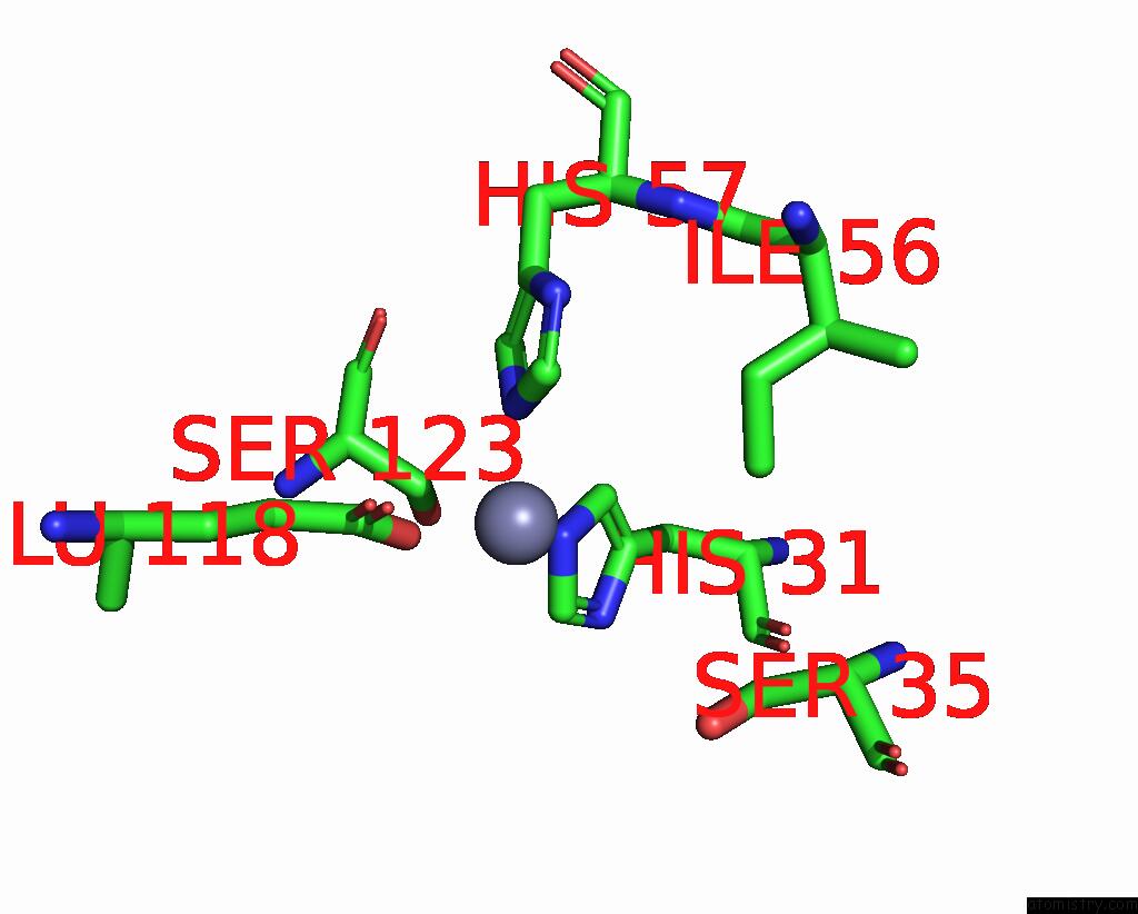
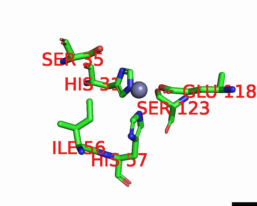
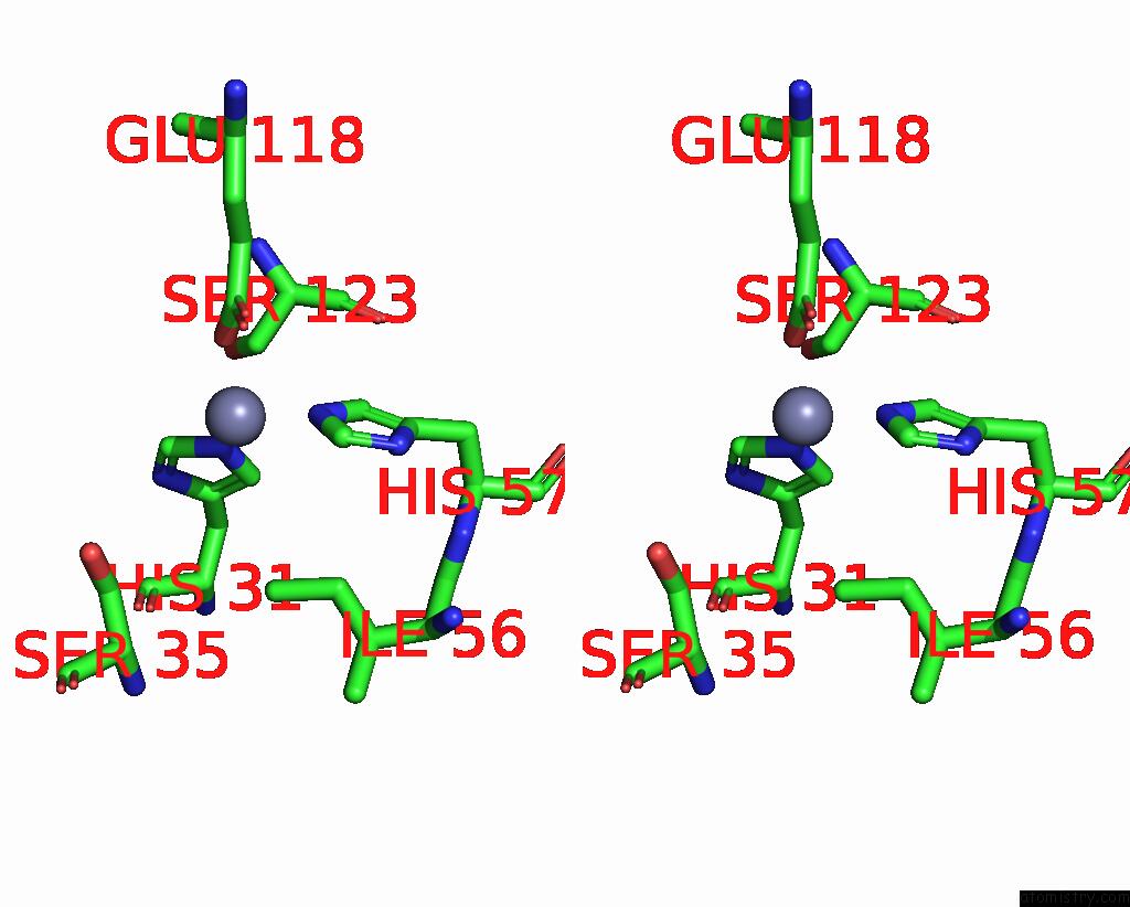
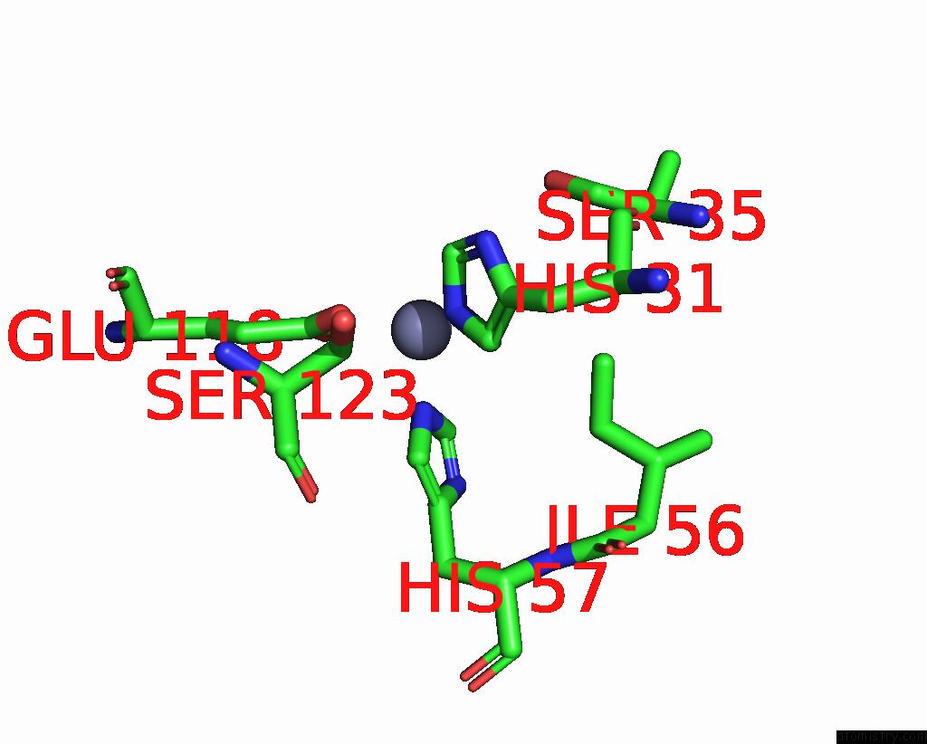
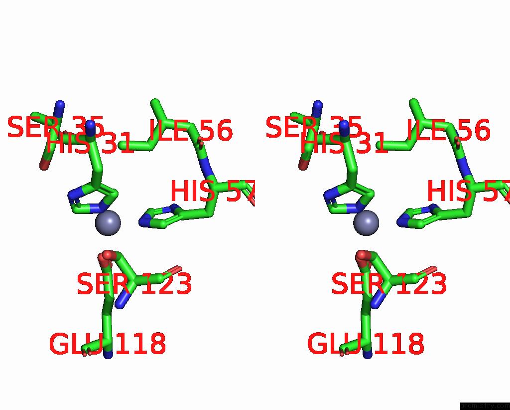
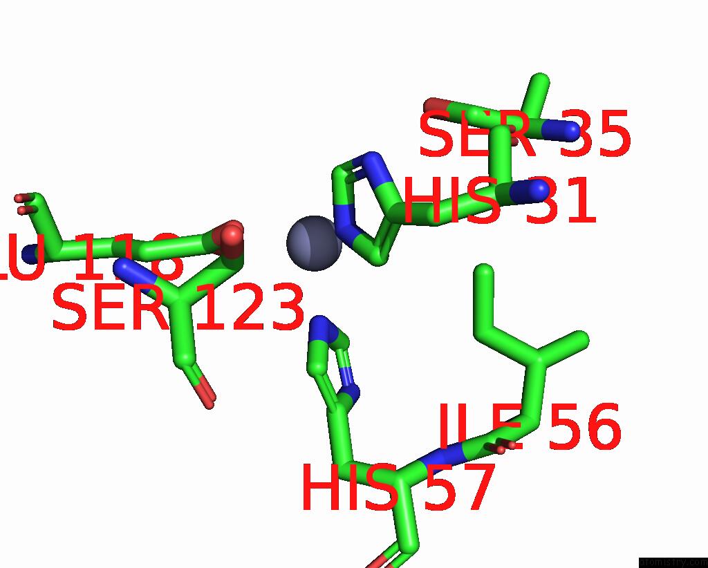
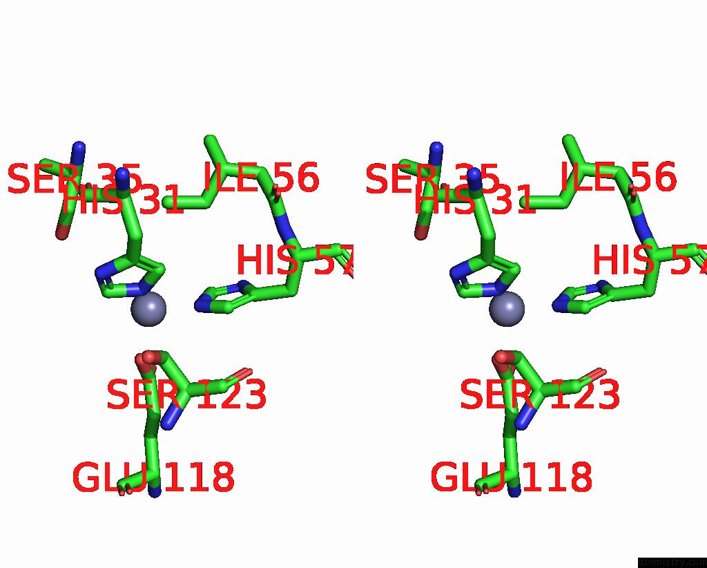
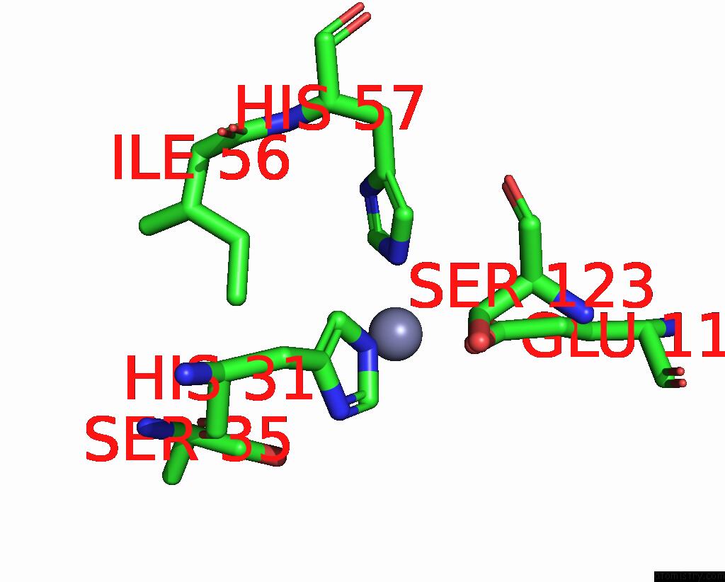
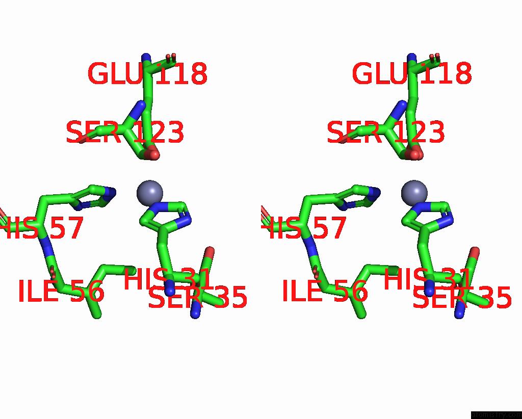
Zinc binding site 1 out of 12 in 1yhu
Go back to
Zinc binding site 1 out
of 12 in the Crystal Structure of Riftia Pachyptila C1 Hemoglobin Reveals Novel Assembly of 24 Subunits.
Mono view
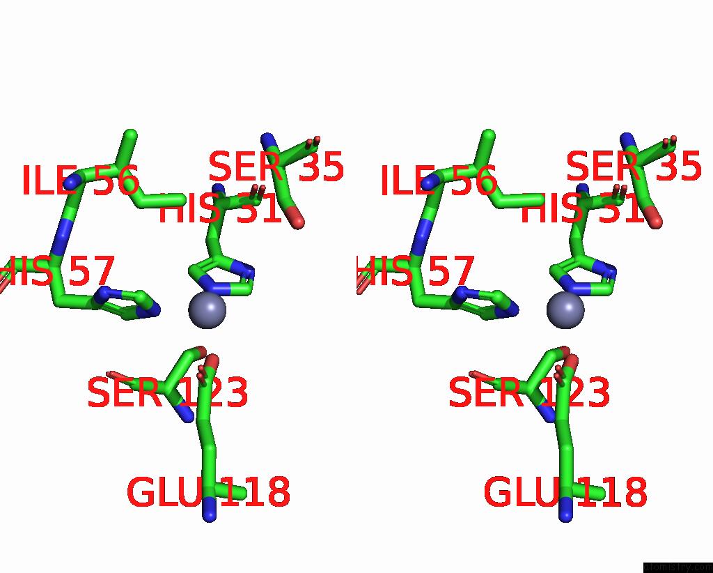
Stereo pair view
Mono view
Stereo pair view
A full contact list of Zinc with other atoms in the Zn binding
site number 1 of Crystal Structure of Riftia Pachyptila C1 Hemoglobin Reveals Novel Assembly of 24 Subunits. within 5.0Å range:
|
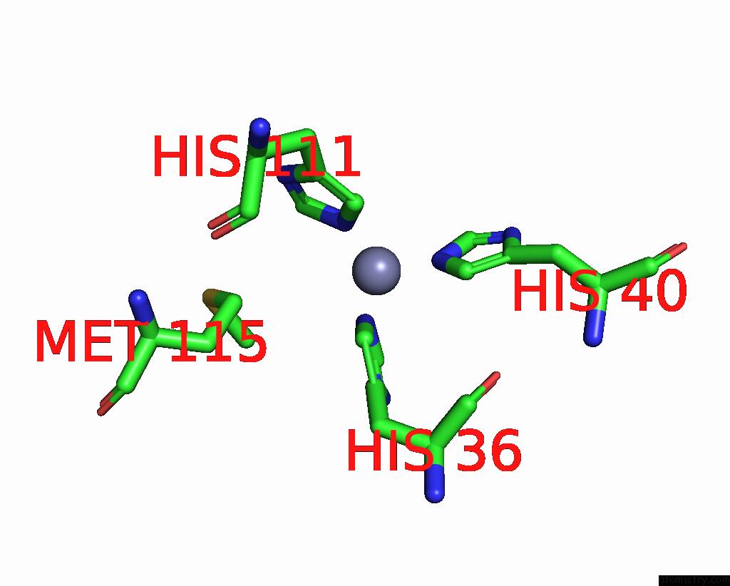
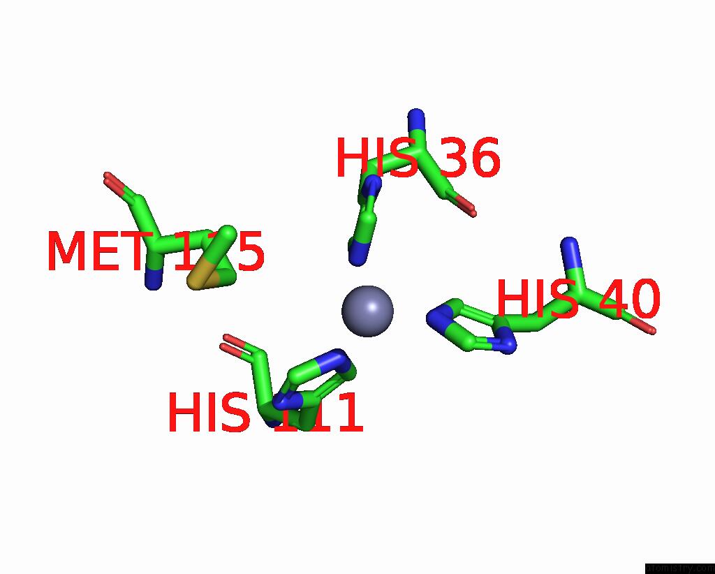
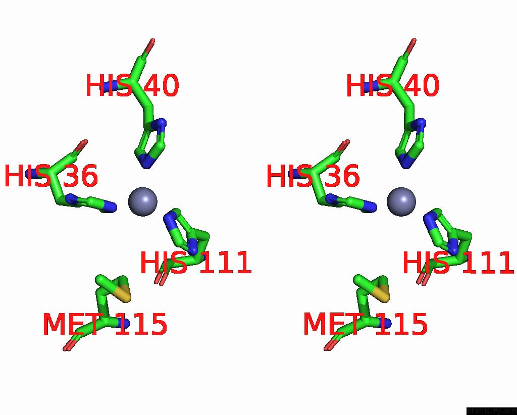
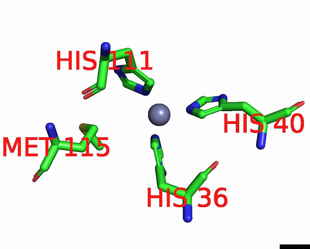
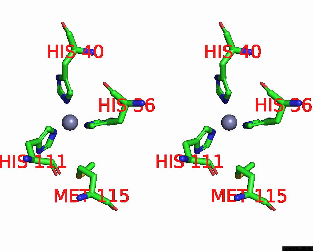
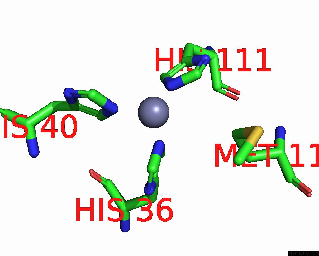
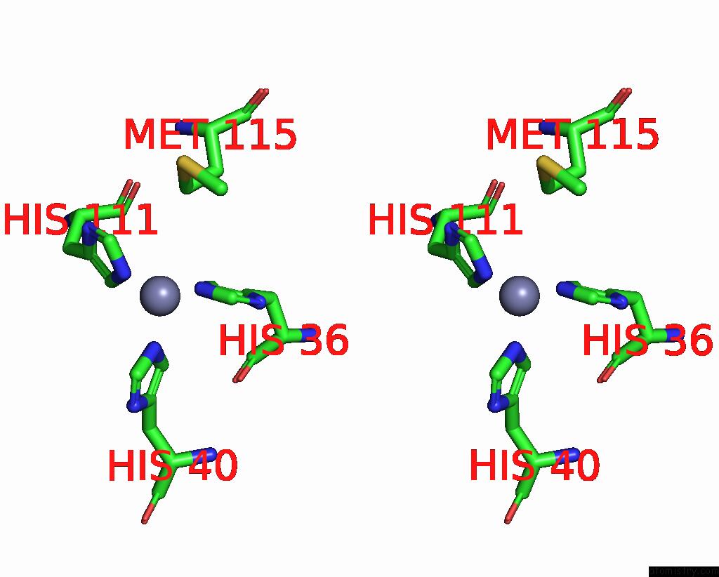
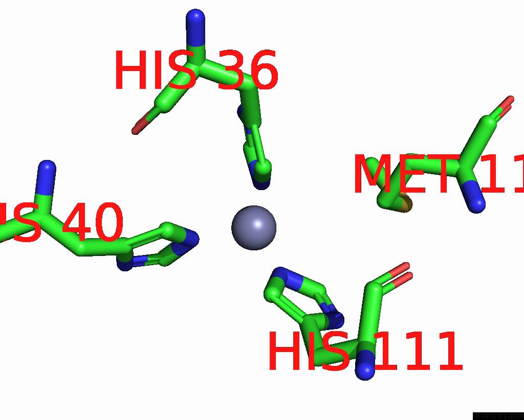
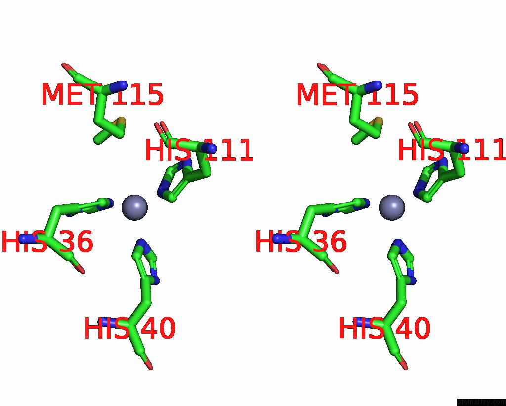
Zinc binding site 2 out of 12 in 1yhu
Go back to
Zinc binding site 2 out
of 12 in the Crystal Structure of Riftia Pachyptila C1 Hemoglobin Reveals Novel Assembly of 24 Subunits.
Mono view

Stereo pair view
Mono view
Stereo pair view
A full contact list of Zinc with other atoms in the Zn binding
site number 2 of Crystal Structure of Riftia Pachyptila C1 Hemoglobin Reveals Novel Assembly of 24 Subunits. within 5.0Å range:
|
Zinc binding site 3 out of 12 in 1yhu
Go back to
Zinc binding site 3 out
of 12 in the Crystal Structure of Riftia Pachyptila C1 Hemoglobin Reveals Novel Assembly of 24 Subunits.
Mono view
Stereo pair view
Mono view
Stereo pair view
A full contact list of Zinc with other atoms in the Zn binding
site number 3 of Crystal Structure of Riftia Pachyptila C1 Hemoglobin Reveals Novel Assembly of 24 Subunits. within 5.0Å range:
|
Zinc binding site 4 out of 12 in 1yhu
Go back to
Zinc binding site 4 out
of 12 in the Crystal Structure of Riftia Pachyptila C1 Hemoglobin Reveals Novel Assembly of 24 Subunits.
Mono view
Stereo pair view
Mono view
Stereo pair view
A full contact list of Zinc with other atoms in the Zn binding
site number 4 of Crystal Structure of Riftia Pachyptila C1 Hemoglobin Reveals Novel Assembly of 24 Subunits. within 5.0Å range:
|
Zinc binding site 5 out of 12 in 1yhu
Go back to
Zinc binding site 5 out
of 12 in the Crystal Structure of Riftia Pachyptila C1 Hemoglobin Reveals Novel Assembly of 24 Subunits.
Mono view
Stereo pair view
Mono view
Stereo pair view
A full contact list of Zinc with other atoms in the Zn binding
site number 5 of Crystal Structure of Riftia Pachyptila C1 Hemoglobin Reveals Novel Assembly of 24 Subunits. within 5.0Å range:
|
Zinc binding site 6 out of 12 in 1yhu
Go back to
Zinc binding site 6 out
of 12 in the Crystal Structure of Riftia Pachyptila C1 Hemoglobin Reveals Novel Assembly of 24 Subunits.
Mono view
Stereo pair view
Mono view
Stereo pair view
A full contact list of Zinc with other atoms in the Zn binding
site number 6 of Crystal Structure of Riftia Pachyptila C1 Hemoglobin Reveals Novel Assembly of 24 Subunits. within 5.0Å range:
|
Zinc binding site 7 out of 12 in 1yhu
Go back to
Zinc binding site 7 out
of 12 in the Crystal Structure of Riftia Pachyptila C1 Hemoglobin Reveals Novel Assembly of 24 Subunits.
Mono view
Stereo pair view
Mono view
Stereo pair view
A full contact list of Zinc with other atoms in the Zn binding
site number 7 of Crystal Structure of Riftia Pachyptila C1 Hemoglobin Reveals Novel Assembly of 24 Subunits. within 5.0Å range:
|
Zinc binding site 8 out of 12 in 1yhu
Go back to
Zinc binding site 8 out
of 12 in the Crystal Structure of Riftia Pachyptila C1 Hemoglobin Reveals Novel Assembly of 24 Subunits.
Mono view
Stereo pair view
Mono view
Stereo pair view
A full contact list of Zinc with other atoms in the Zn binding
site number 8 of Crystal Structure of Riftia Pachyptila C1 Hemoglobin Reveals Novel Assembly of 24 Subunits. within 5.0Å range:
|
Zinc binding site 9 out of 12 in 1yhu
Go back to
Zinc binding site 9 out
of 12 in the Crystal Structure of Riftia Pachyptila C1 Hemoglobin Reveals Novel Assembly of 24 Subunits.
Mono view
Stereo pair view
Mono view
Stereo pair view
A full contact list of Zinc with other atoms in the Zn binding
site number 9 of Crystal Structure of Riftia Pachyptila C1 Hemoglobin Reveals Novel Assembly of 24 Subunits. within 5.0Å range:
|
Zinc binding site 10 out of 12 in 1yhu
Go back to
Zinc binding site 10 out
of 12 in the Crystal Structure of Riftia Pachyptila C1 Hemoglobin Reveals Novel Assembly of 24 Subunits.
Mono view
Stereo pair view
Mono view
Stereo pair view
A full contact list of Zinc with other atoms in the Zn binding
site number 10 of Crystal Structure of Riftia Pachyptila C1 Hemoglobin Reveals Novel Assembly of 24 Subunits. within 5.0Å range:
|
Reference:
J.F.Flores,
C.R.Fisher,
S.L.Carney,
B.N.Green,
J.K.Freytag,
S.W.Schaeffer,
W.E.Royer Jr.
Sulfide Binding Is Mediated By Zinc Ions Discovered in the Crystal Structure of A Hydrothermal Vent Tubeworm Hemoglobin. Proc.Natl.Acad.Sci.Usa V. 102 2713 2005.
ISSN: ISSN 0027-8424
PubMed: 15710902
DOI: 10.1073/PNAS.0407455102
Page generated: Wed Oct 16 20:57:21 2024
ISSN: ISSN 0027-8424
PubMed: 15710902
DOI: 10.1073/PNAS.0407455102
Last articles
As in 3A6FAs in 3CFS
As in 3CAO
As in 3BFD
As in 3A6E
As in 3BWW
As in 3BFT
As in 2Z3J
As in 3AAL
As in 2Z3I