Zinc »
PDB 1y8q-1ykf »
1yfh »
Zinc in PDB 1yfh: Wt Human O6-Alkylguanine-Dna Alkyltransferase Bound to Dna Containing An Alkylated Cytosine
Enzymatic activity of Wt Human O6-Alkylguanine-Dna Alkyltransferase Bound to Dna Containing An Alkylated Cytosine
All present enzymatic activity of Wt Human O6-Alkylguanine-Dna Alkyltransferase Bound to Dna Containing An Alkylated Cytosine:
2.1.1.63;
2.1.1.63;
Protein crystallography data
The structure of Wt Human O6-Alkylguanine-Dna Alkyltransferase Bound to Dna Containing An Alkylated Cytosine, PDB code: 1yfh
was solved by
E.M.Duguid,
P.A.Rice,
C.He,
with X-Ray Crystallography technique. A brief refinement statistics is given in the table below:
| Resolution Low / High (Å) | 29.17 / 3.01 |
| Space group | P 1 21 1 |
| Cell size a, b, c (Å), α, β, γ (°) | 58.150, 102.710, 87.940, 90.00, 106.77, 90.00 |
| R / Rfree (%) | 24.6 / 28.6 |
Zinc Binding Sites:
The binding sites of Zinc atom in the Wt Human O6-Alkylguanine-Dna Alkyltransferase Bound to Dna Containing An Alkylated Cytosine
(pdb code 1yfh). This binding sites where shown within
5.0 Angstroms radius around Zinc atom.
In total 3 binding sites of Zinc where determined in the Wt Human O6-Alkylguanine-Dna Alkyltransferase Bound to Dna Containing An Alkylated Cytosine, PDB code: 1yfh:
Jump to Zinc binding site number: 1; 2; 3;
In total 3 binding sites of Zinc where determined in the Wt Human O6-Alkylguanine-Dna Alkyltransferase Bound to Dna Containing An Alkylated Cytosine, PDB code: 1yfh:
Jump to Zinc binding site number: 1; 2; 3;
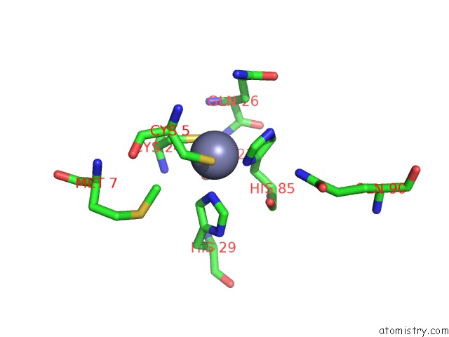
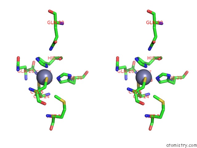
Zinc binding site 1 out of 3 in 1yfh
Go back to
Zinc binding site 1 out
of 3 in the Wt Human O6-Alkylguanine-Dna Alkyltransferase Bound to Dna Containing An Alkylated Cytosine
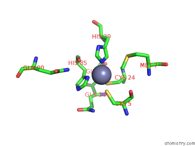
Mono view
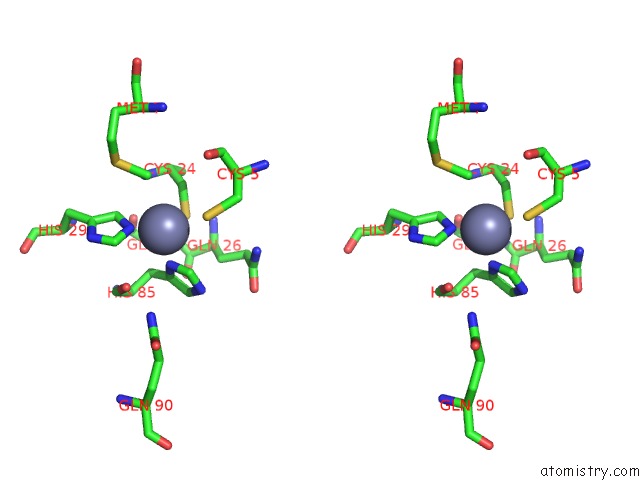
Stereo pair view
Mono view
Stereo pair view
A full contact list of Zinc with other atoms in the Zn binding
site number 1 of Wt Human O6-Alkylguanine-Dna Alkyltransferase Bound to Dna Containing An Alkylated Cytosine within 5.0Å range:
|
Zinc binding site 2 out of 3 in 1yfh
Go back to
Zinc binding site 2 out
of 3 in the Wt Human O6-Alkylguanine-Dna Alkyltransferase Bound to Dna Containing An Alkylated Cytosine
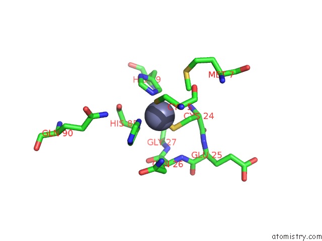
Mono view
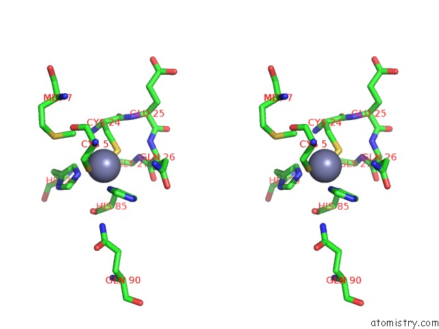
Stereo pair view
Mono view
Stereo pair view
A full contact list of Zinc with other atoms in the Zn binding
site number 2 of Wt Human O6-Alkylguanine-Dna Alkyltransferase Bound to Dna Containing An Alkylated Cytosine within 5.0Å range:
|
Zinc binding site 3 out of 3 in 1yfh
Go back to
Zinc binding site 3 out
of 3 in the Wt Human O6-Alkylguanine-Dna Alkyltransferase Bound to Dna Containing An Alkylated Cytosine
Mono view
Stereo pair view
Mono view
Stereo pair view
A full contact list of Zinc with other atoms in the Zn binding
site number 3 of Wt Human O6-Alkylguanine-Dna Alkyltransferase Bound to Dna Containing An Alkylated Cytosine within 5.0Å range:
|
Reference:
E.M.Duguid,
P.A.Rice,
C.He.
The Structure of the Human Agt Protein Bound to Dna and Its Implications For Damage Detection. J.Mol.Biol. V. 350 657 2005.
ISSN: ISSN 0022-2836
PubMed: 15964013
DOI: 10.1016/J.JMB.2005.05.028
Page generated: Wed Oct 16 20:56:14 2024
ISSN: ISSN 0022-2836
PubMed: 15964013
DOI: 10.1016/J.JMB.2005.05.028
Last articles
Zn in 9MJ5Zn in 9HNW
Zn in 9G0L
Zn in 9FNE
Zn in 9DZN
Zn in 9E0I
Zn in 9D32
Zn in 9DAK
Zn in 8ZXC
Zn in 8ZUF