Zinc »
PDB 1y8q-1ykf »
1yeh »
Zinc in PDB 1yeh: Structure of IGG2A Fab Fragment
Protein crystallography data
The structure of Structure of IGG2A Fab Fragment, PDB code: 1yeh
was solved by
B.Gigant,
M.Knossow,
with X-Ray Crystallography technique. A brief refinement statistics is given in the table below:
| Resolution Low / High (Å) | 7.00 / 2.55 |
| Space group | P 31 2 1 |
| Cell size a, b, c (Å), α, β, γ (°) | 78.470, 78.470, 158.920, 90.00, 90.00, 120.00 |
| R / Rfree (%) | 18.1 / 23.1 |
Zinc Binding Sites:
The binding sites of Zinc atom in the Structure of IGG2A Fab Fragment
(pdb code 1yeh). This binding sites where shown within
5.0 Angstroms radius around Zinc atom.
In total 7 binding sites of Zinc where determined in the Structure of IGG2A Fab Fragment, PDB code: 1yeh:
Jump to Zinc binding site number: 1; 2; 3; 4; 5; 6; 7;
In total 7 binding sites of Zinc where determined in the Structure of IGG2A Fab Fragment, PDB code: 1yeh:
Jump to Zinc binding site number: 1; 2; 3; 4; 5; 6; 7;
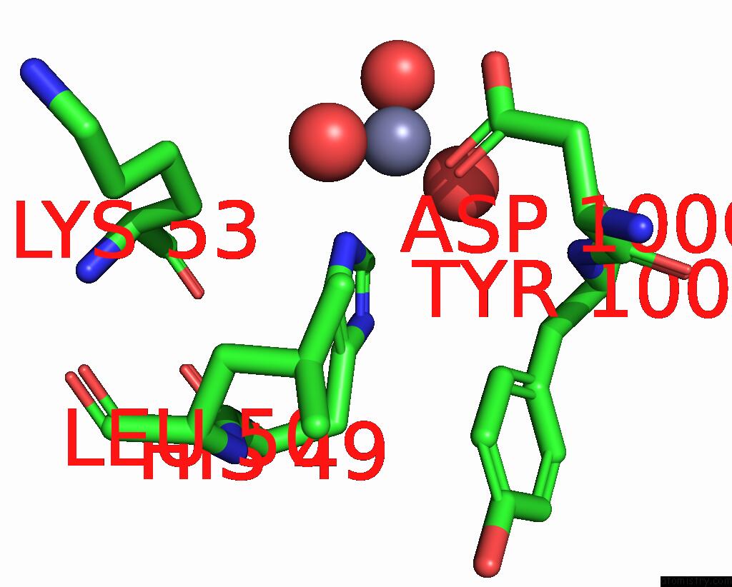
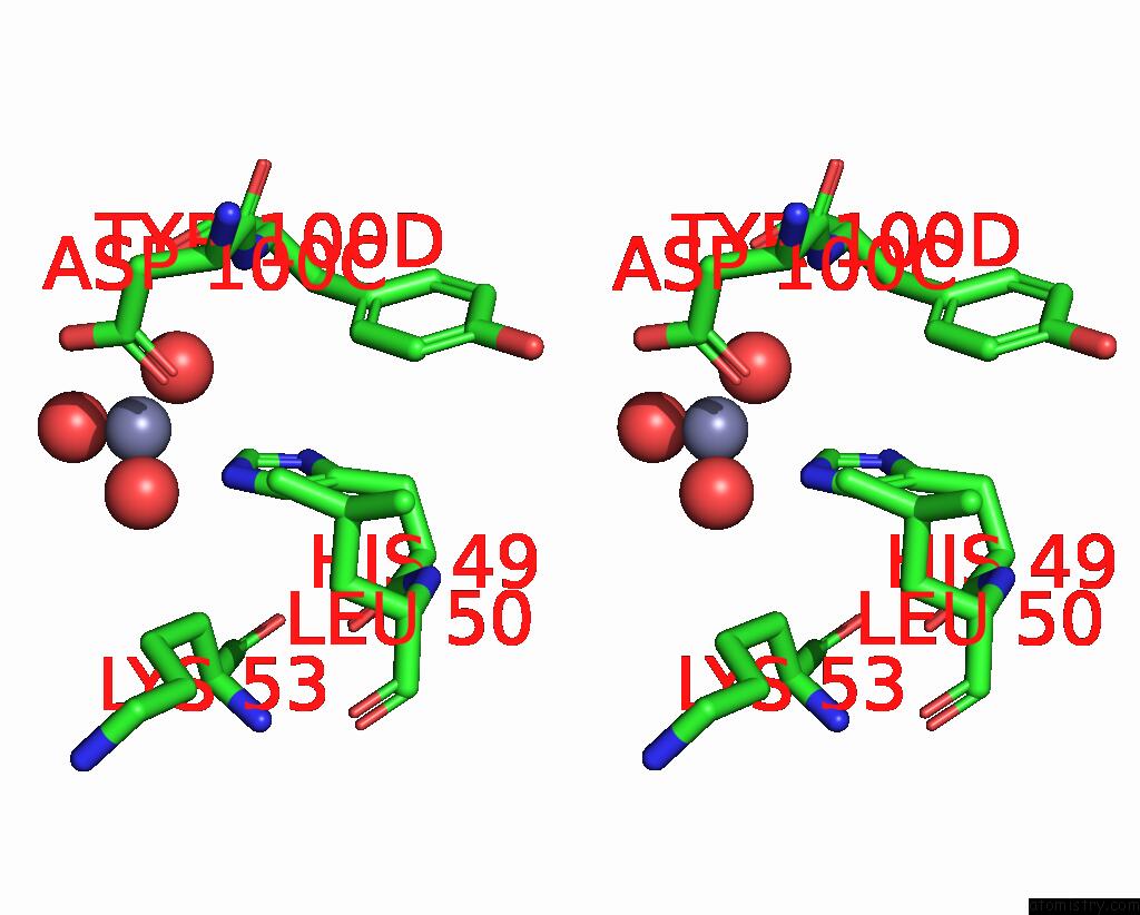
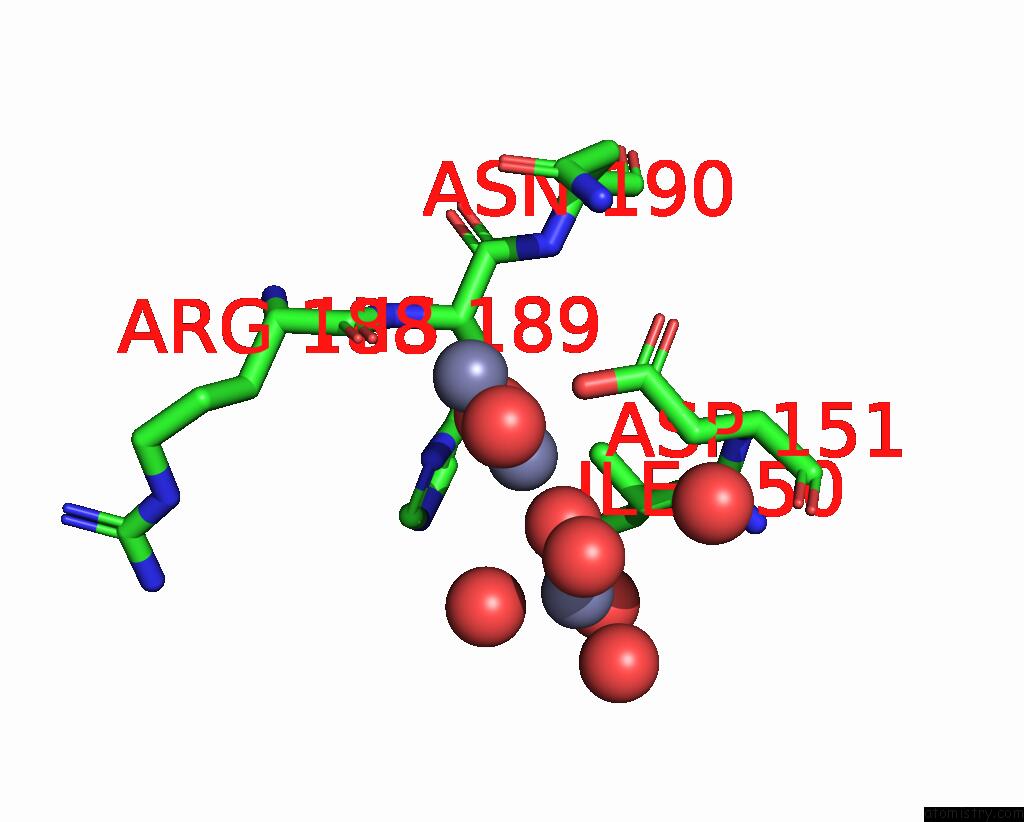
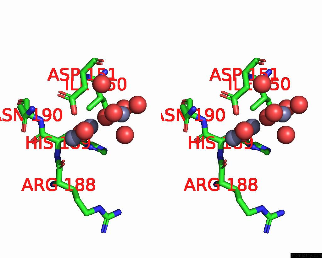
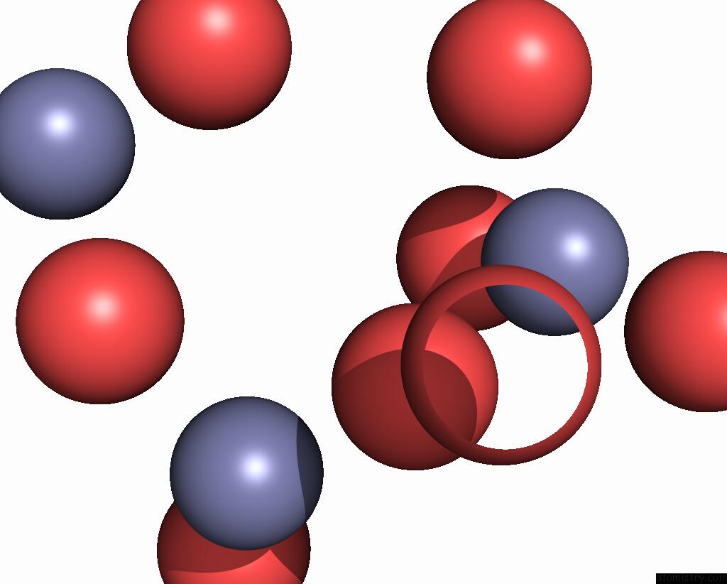
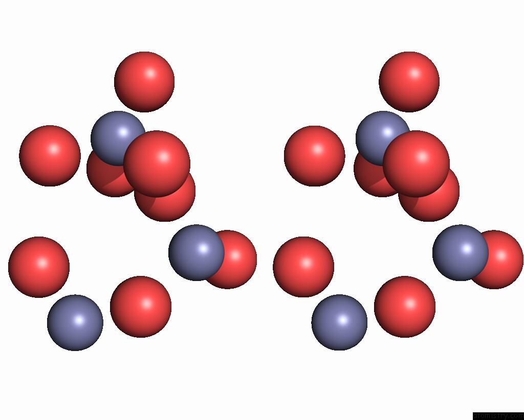
Zinc binding site 1 out of 7 in 1yeh
Go back to
Zinc binding site 1 out
of 7 in the Structure of IGG2A Fab Fragment
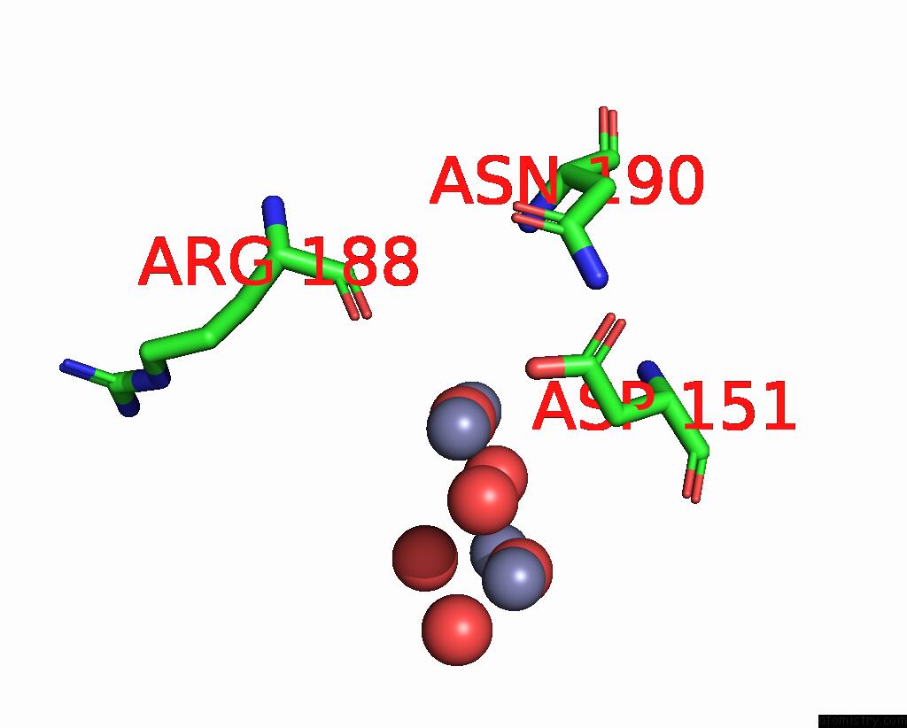
Mono view
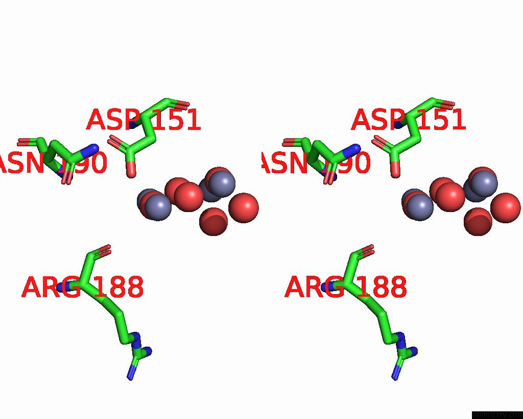
Stereo pair view
Mono view
Stereo pair view
A full contact list of Zinc with other atoms in the Zn binding
site number 1 of Structure of IGG2A Fab Fragment within 5.0Å range:
|
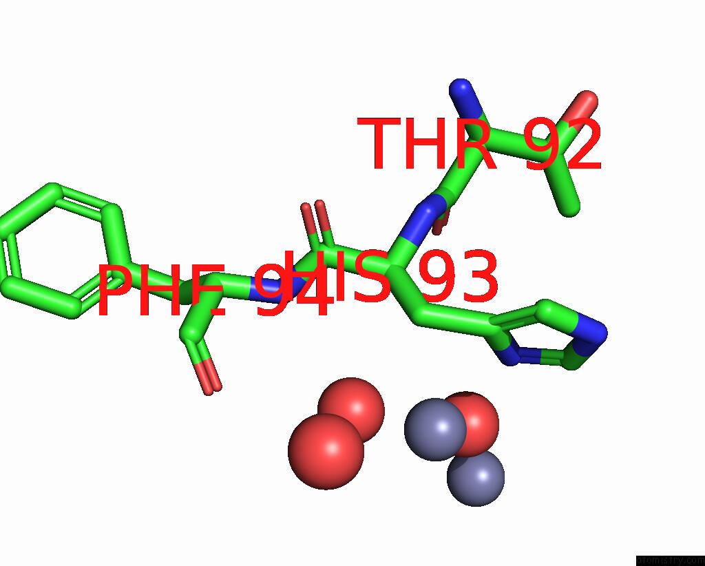
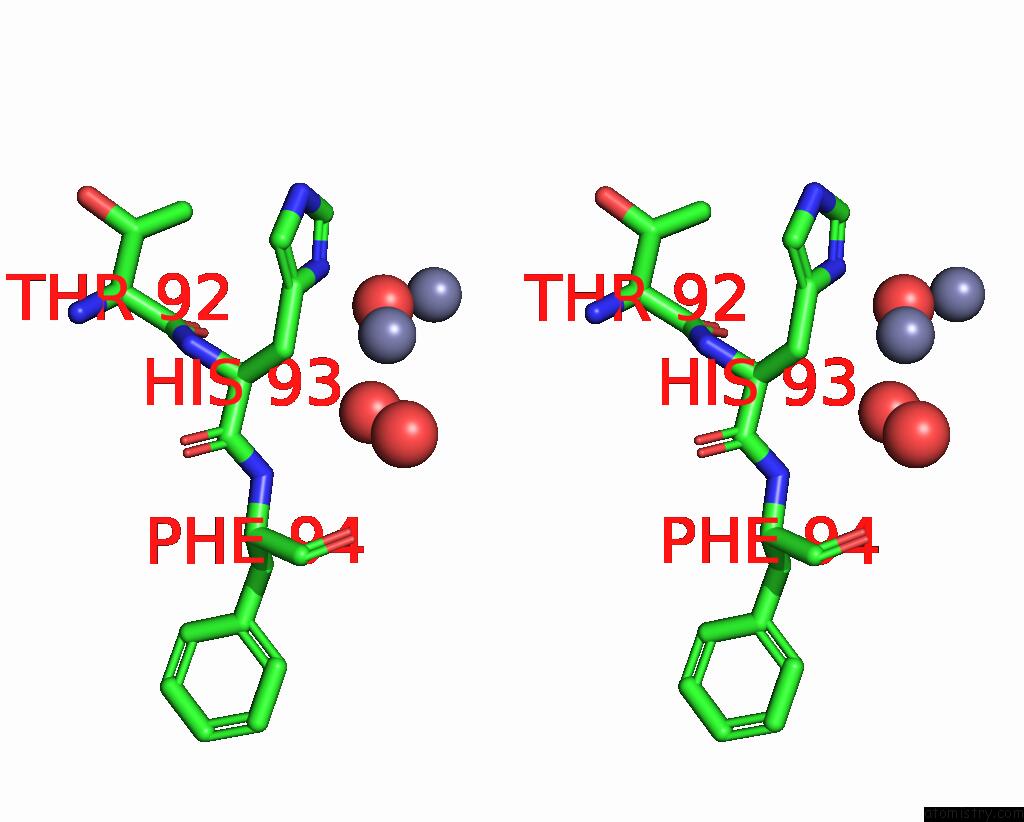
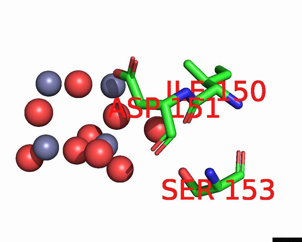
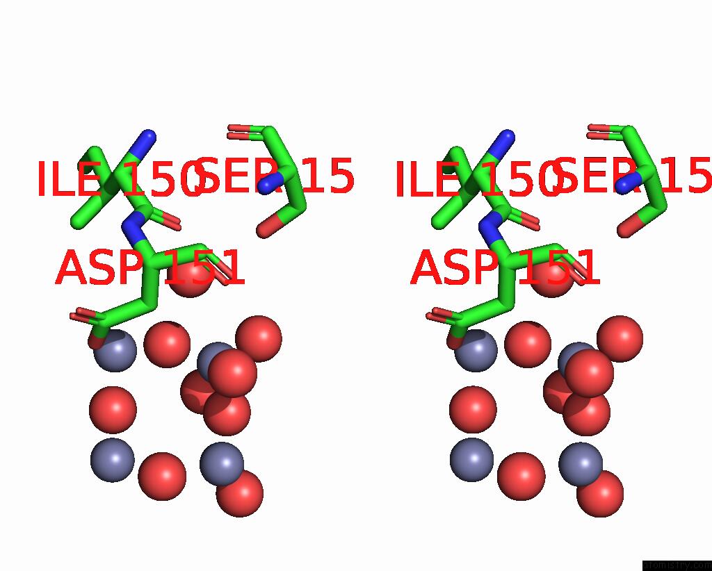
Zinc binding site 2 out of 7 in 1yeh
Go back to
Zinc binding site 2 out
of 7 in the Structure of IGG2A Fab Fragment
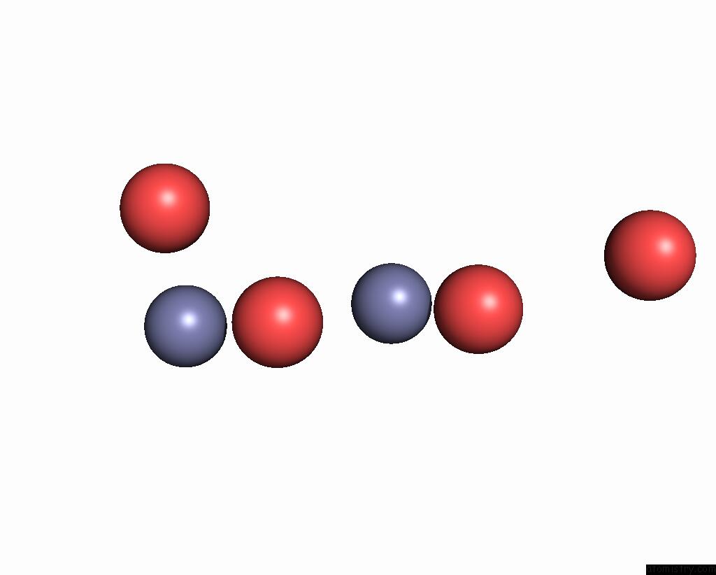
Mono view
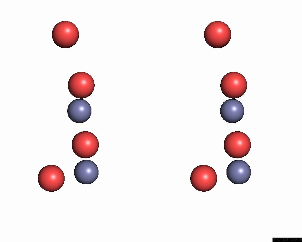
Stereo pair view
Mono view
Stereo pair view
A full contact list of Zinc with other atoms in the Zn binding
site number 2 of Structure of IGG2A Fab Fragment within 5.0Å range:
|
Zinc binding site 3 out of 7 in 1yeh
Go back to
Zinc binding site 3 out
of 7 in the Structure of IGG2A Fab Fragment
Mono view
Stereo pair view
Mono view
Stereo pair view
A full contact list of Zinc with other atoms in the Zn binding
site number 3 of Structure of IGG2A Fab Fragment within 5.0Å range:
|
Zinc binding site 4 out of 7 in 1yeh
Go back to
Zinc binding site 4 out
of 7 in the Structure of IGG2A Fab Fragment
Mono view
Stereo pair view
Mono view
Stereo pair view
A full contact list of Zinc with other atoms in the Zn binding
site number 4 of Structure of IGG2A Fab Fragment within 5.0Å range:
|
Zinc binding site 5 out of 7 in 1yeh
Go back to
Zinc binding site 5 out
of 7 in the Structure of IGG2A Fab Fragment
Mono view
Stereo pair view
Mono view
Stereo pair view
A full contact list of Zinc with other atoms in the Zn binding
site number 5 of Structure of IGG2A Fab Fragment within 5.0Å range:
|
Zinc binding site 6 out of 7 in 1yeh
Go back to
Zinc binding site 6 out
of 7 in the Structure of IGG2A Fab Fragment
Mono view
Stereo pair view
Mono view
Stereo pair view
A full contact list of Zinc with other atoms in the Zn binding
site number 6 of Structure of IGG2A Fab Fragment within 5.0Å range:
|
Zinc binding site 7 out of 7 in 1yeh
Go back to
Zinc binding site 7 out
of 7 in the Structure of IGG2A Fab Fragment
Mono view
Stereo pair view
Mono view
Stereo pair view
A full contact list of Zinc with other atoms in the Zn binding
site number 7 of Structure of IGG2A Fab Fragment within 5.0Å range:
|
Reference:
B.Gigant,
J.B.Charbonnier,
Z.Eshhar,
B.S.Green,
M.Knossow.
X-Ray Structures of A Hydrolytic Antibody and of Complexes Elucidate Catalytic Pathway From Substrate Binding and Transition State Stabilization Through Water Attack and Product Release. Proc.Natl.Acad.Sci.Usa V. 94 7857 1997.
ISSN: ISSN 0027-8424
PubMed: 9223277
DOI: 10.1073/PNAS.94.15.7857
Page generated: Wed Oct 16 20:54:03 2024
ISSN: ISSN 0027-8424
PubMed: 9223277
DOI: 10.1073/PNAS.94.15.7857
Last articles
Al in 1OH9Al in 1L7N
Al in 1N6K
Al in 1MND
Al in 1K5G
Al in 1KDN
Al in 1L3R
Al in 1KHJ
Al in 1KH5
Al in 1H8E