Zinc »
PDB 1y8q-1ykf »
1ydc »
Zinc in PDB 1ydc: Structural Basis of Inhibitor Affinity to Variants of Human Carbonic Anhydrase II
Enzymatic activity of Structural Basis of Inhibitor Affinity to Variants of Human Carbonic Anhydrase II
All present enzymatic activity of Structural Basis of Inhibitor Affinity to Variants of Human Carbonic Anhydrase II:
4.2.1.1;
4.2.1.1;
Protein crystallography data
The structure of Structural Basis of Inhibitor Affinity to Variants of Human Carbonic Anhydrase II, PDB code: 1ydc
was solved by
S.K.Nair,
D.W.Christianson,
with X-Ray Crystallography technique. A brief refinement statistics is given in the table below:
| Resolution Low / High (Å) | 6.50 / 1.95 |
| Space group | P 1 21 1 |
| Cell size a, b, c (Å), α, β, γ (°) | 42.700, 41.700, 73.000, 90.00, 104.60, 90.00 |
| R / Rfree (%) | 16.5 / n/a |
Other elements in 1ydc:
The structure of Structural Basis of Inhibitor Affinity to Variants of Human Carbonic Anhydrase II also contains other interesting chemical elements:
| Mercury | (Hg) | 1 atom |
Zinc Binding Sites:
The binding sites of Zinc atom in the Structural Basis of Inhibitor Affinity to Variants of Human Carbonic Anhydrase II
(pdb code 1ydc). This binding sites where shown within
5.0 Angstroms radius around Zinc atom.
In total only one binding site of Zinc was determined in the Structural Basis of Inhibitor Affinity to Variants of Human Carbonic Anhydrase II, PDB code: 1ydc:
In total only one binding site of Zinc was determined in the Structural Basis of Inhibitor Affinity to Variants of Human Carbonic Anhydrase II, PDB code: 1ydc:
Zinc binding site 1 out of 1 in 1ydc
Go back to
Zinc binding site 1 out
of 1 in the Structural Basis of Inhibitor Affinity to Variants of Human Carbonic Anhydrase II
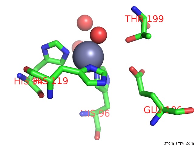
Mono view
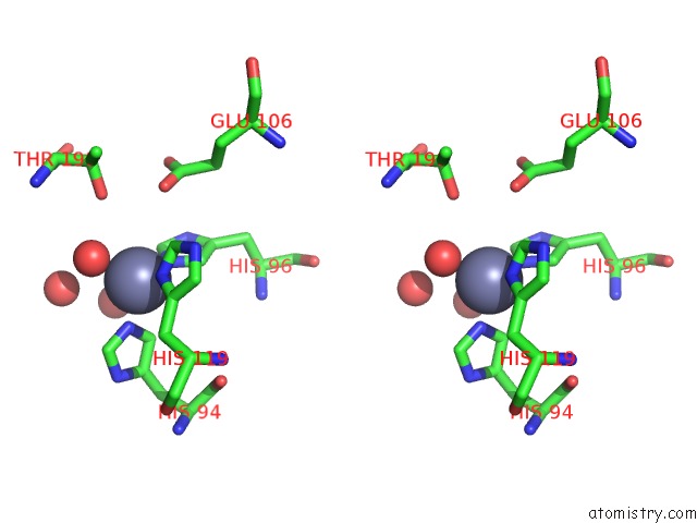
Stereo pair view
Mono view
Stereo pair view
A full contact list of Zinc with other atoms in the Zn binding
site number 1 of Structural Basis of Inhibitor Affinity to Variants of Human Carbonic Anhydrase II within 5.0Å range:
|
Reference:
S.K.Nair,
J.F.Krebs,
D.W.Christianson,
C.A.Fierke.
Structural Basis of Inhibitor Affinity to Variants of Human Carbonic Anhydrase II. Biochemistry V. 34 3981 1995.
ISSN: ISSN 0006-2960
PubMed: 7696263
DOI: 10.1021/BI00012A016
Page generated: Wed Oct 16 20:52:28 2024
ISSN: ISSN 0006-2960
PubMed: 7696263
DOI: 10.1021/BI00012A016
Last articles
Zn in 9MJ5Zn in 9HNW
Zn in 9G0L
Zn in 9FNE
Zn in 9DZN
Zn in 9E0I
Zn in 9D32
Zn in 9DAK
Zn in 8ZXC
Zn in 8ZUF