Zinc »
PDB 1y8q-1ykf »
1y93 »
Zinc in PDB 1y93: Crystal Structure of the Catalytic Domain of Human MMP12 Complexed with Acetohydroxamic Acid at Atomic Resolution
Enzymatic activity of Crystal Structure of the Catalytic Domain of Human MMP12 Complexed with Acetohydroxamic Acid at Atomic Resolution
All present enzymatic activity of Crystal Structure of the Catalytic Domain of Human MMP12 Complexed with Acetohydroxamic Acid at Atomic Resolution:
3.4.24.65;
3.4.24.65;
Protein crystallography data
The structure of Crystal Structure of the Catalytic Domain of Human MMP12 Complexed with Acetohydroxamic Acid at Atomic Resolution, PDB code: 1y93
was solved by
I.Bertini,
V.Calderone,
M.Cosenza,
M.Fragai,
Y.-M.Lee,
C.Luchinat,
S.Mangani,
B.Terni,
P.Turano,
with X-Ray Crystallography technique. A brief refinement statistics is given in the table below:
| Resolution Low / High (Å) | 13.99 / 1.03 |
| Space group | C 1 2 1 |
| Cell size a, b, c (Å), α, β, γ (°) | 50.913, 59.552, 53.497, 90.00, 115.14, 90.00 |
| R / Rfree (%) | 15.6 / 16.8 |
Other elements in 1y93:
The structure of Crystal Structure of the Catalytic Domain of Human MMP12 Complexed with Acetohydroxamic Acid at Atomic Resolution also contains other interesting chemical elements:
| Calcium | (Ca) | 3 atoms |
Zinc Binding Sites:
The binding sites of Zinc atom in the Crystal Structure of the Catalytic Domain of Human MMP12 Complexed with Acetohydroxamic Acid at Atomic Resolution
(pdb code 1y93). This binding sites where shown within
5.0 Angstroms radius around Zinc atom.
In total 2 binding sites of Zinc where determined in the Crystal Structure of the Catalytic Domain of Human MMP12 Complexed with Acetohydroxamic Acid at Atomic Resolution, PDB code: 1y93:
Jump to Zinc binding site number: 1; 2;
In total 2 binding sites of Zinc where determined in the Crystal Structure of the Catalytic Domain of Human MMP12 Complexed with Acetohydroxamic Acid at Atomic Resolution, PDB code: 1y93:
Jump to Zinc binding site number: 1; 2;
Zinc binding site 1 out of 2 in 1y93
Go back to
Zinc binding site 1 out
of 2 in the Crystal Structure of the Catalytic Domain of Human MMP12 Complexed with Acetohydroxamic Acid at Atomic Resolution
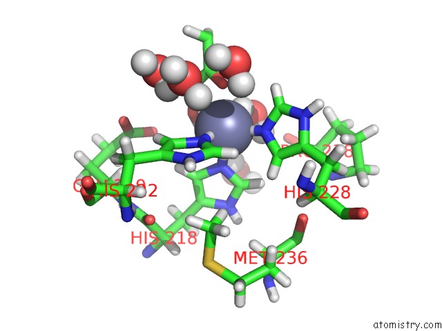
Mono view
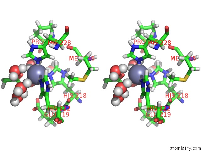
Stereo pair view
Mono view
Stereo pair view
A full contact list of Zinc with other atoms in the Zn binding
site number 1 of Crystal Structure of the Catalytic Domain of Human MMP12 Complexed with Acetohydroxamic Acid at Atomic Resolution within 5.0Å range:
|
Zinc binding site 2 out of 2 in 1y93
Go back to
Zinc binding site 2 out
of 2 in the Crystal Structure of the Catalytic Domain of Human MMP12 Complexed with Acetohydroxamic Acid at Atomic Resolution
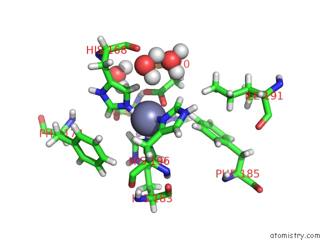
Mono view
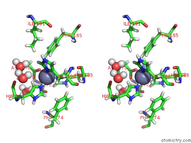
Stereo pair view
Mono view
Stereo pair view
A full contact list of Zinc with other atoms in the Zn binding
site number 2 of Crystal Structure of the Catalytic Domain of Human MMP12 Complexed with Acetohydroxamic Acid at Atomic Resolution within 5.0Å range:
|
Reference:
I.Bertini,
V.Calderone,
M.Cosenza,
M.Fragai,
Y.-M.Lee,
C.Luchinat,
S.Mangani,
B.Terni,
P.Turano.
Conformational Variability of Matrix Metalloproteinases: Beyond A Single 3D Structure Proc.Natl.Acad.Sci.Usa V. 102 5334 2005.
ISSN: ISSN 0027-8424
PubMed: 15809432
DOI: 10.1073/PNAS.0407106102
Page generated: Wed Oct 16 20:48:23 2024
ISSN: ISSN 0027-8424
PubMed: 15809432
DOI: 10.1073/PNAS.0407106102
Last articles
Zn in 9MJ5Zn in 9HNW
Zn in 9G0L
Zn in 9FNE
Zn in 9DZN
Zn in 9E0I
Zn in 9D32
Zn in 9DAK
Zn in 8ZXC
Zn in 8ZUF