Zinc »
PDB 1xv2-1y8j »
1xwy »
Zinc in PDB 1xwy: Crystal Structure of Tatd Deoxyribonuclease From Escherichia Coli K12 at 2.0 A Resolution
Protein crystallography data
The structure of Crystal Structure of Tatd Deoxyribonuclease From Escherichia Coli K12 at 2.0 A Resolution, PDB code: 1xwy
was solved by
V.N.Malashkevich,
D.F.Xiang,
F.M.Raushel,
S.C.Almo,
S.K.Burley,
New Yorksgx Research Center For Structural Genomics (Nysgxrc),
with X-Ray Crystallography technique. A brief refinement statistics is given in the table below:
| Resolution Low / High (Å) | 19.93 / 2.00 |
| Space group | P 41 |
| Cell size a, b, c (Å), α, β, γ (°) | 73.885, 73.885, 67.824, 90.00, 90.00, 90.00 |
| R / Rfree (%) | 17.1 / 21.6 |
Zinc Binding Sites:
The binding sites of Zinc atom in the Crystal Structure of Tatd Deoxyribonuclease From Escherichia Coli K12 at 2.0 A Resolution
(pdb code 1xwy). This binding sites where shown within
5.0 Angstroms radius around Zinc atom.
In total only one binding site of Zinc was determined in the Crystal Structure of Tatd Deoxyribonuclease From Escherichia Coli K12 at 2.0 A Resolution, PDB code: 1xwy:
In total only one binding site of Zinc was determined in the Crystal Structure of Tatd Deoxyribonuclease From Escherichia Coli K12 at 2.0 A Resolution, PDB code: 1xwy:
Zinc binding site 1 out of 1 in 1xwy
Go back to
Zinc binding site 1 out
of 1 in the Crystal Structure of Tatd Deoxyribonuclease From Escherichia Coli K12 at 2.0 A Resolution
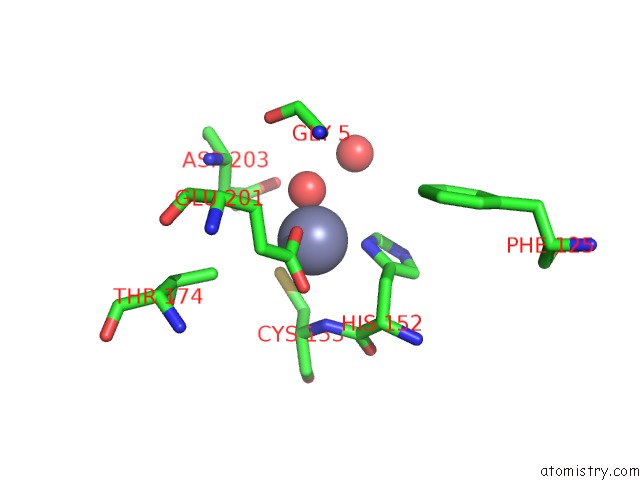
Mono view
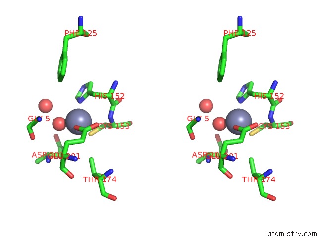
Stereo pair view
Mono view
Stereo pair view
A full contact list of Zinc with other atoms in the Zn binding
site number 1 of Crystal Structure of Tatd Deoxyribonuclease From Escherichia Coli K12 at 2.0 A Resolution within 5.0Å range:
|
Reference:
V.N.Malashkevich,
D.F.Xiang,
F.M.Raushel,
S.C.Almo.
Crystal Structure of Tatd Dnase From Escherichia Coli at 2.0 A Resolution To Be Published.
Page generated: Wed Aug 20 00:27:12 2025
Last articles
Zn in 2GZGZn in 2GZF
Zn in 2GZE
Zn in 2GVI
Zn in 2GYK
Zn in 2GWN
Zn in 2GWG
Zn in 2GVF
Zn in 2GVM
Zn in 2GU2