Zinc »
PDB 1xmo-1xur »
1xtl »
Zinc in PDB 1xtl: Crystal Structure of P104H Mutant of Sod-Like Protein From Bacillus Subtilis.
Protein crystallography data
The structure of Crystal Structure of P104H Mutant of Sod-Like Protein From Bacillus Subtilis., PDB code: 1xtl
was solved by
V.Calderone,
S.Mangani,
L.Banci,
M.Benvenuti,
I.Bertini,
M.S.Viezzoli,
A.Fantoni,
with X-Ray Crystallography technique. A brief refinement statistics is given in the table below:
| Resolution Low / High (Å) | 38.92 / 2.00 |
| Space group | P 1 |
| Cell size a, b, c (Å), α, β, γ (°) | 52.147, 56.586, 59.118, 78.24, 89.91, 85.47 |
| R / Rfree (%) | 23.2 / 29.9 |
Other elements in 1xtl:
The structure of Crystal Structure of P104H Mutant of Sod-Like Protein From Bacillus Subtilis. also contains other interesting chemical elements:
| Copper | (Cu) | 4 atoms |
Zinc Binding Sites:
The binding sites of Zinc atom in the Crystal Structure of P104H Mutant of Sod-Like Protein From Bacillus Subtilis.
(pdb code 1xtl). This binding sites where shown within
5.0 Angstroms radius around Zinc atom.
In total 10 binding sites of Zinc where determined in the Crystal Structure of P104H Mutant of Sod-Like Protein From Bacillus Subtilis., PDB code: 1xtl:
Jump to Zinc binding site number: 1; 2; 3; 4; 5; 6; 7; 8; 9; 10;
In total 10 binding sites of Zinc where determined in the Crystal Structure of P104H Mutant of Sod-Like Protein From Bacillus Subtilis., PDB code: 1xtl:
Jump to Zinc binding site number: 1; 2; 3; 4; 5; 6; 7; 8; 9; 10;
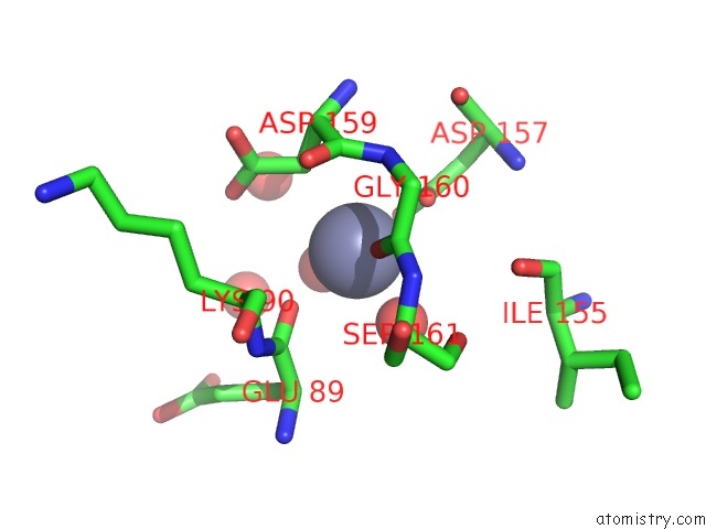
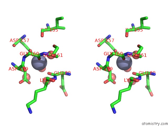
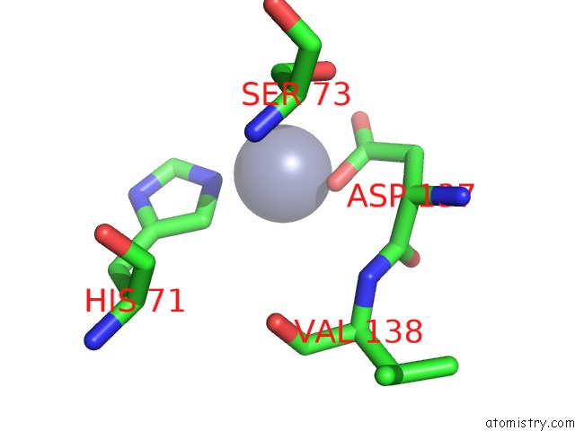
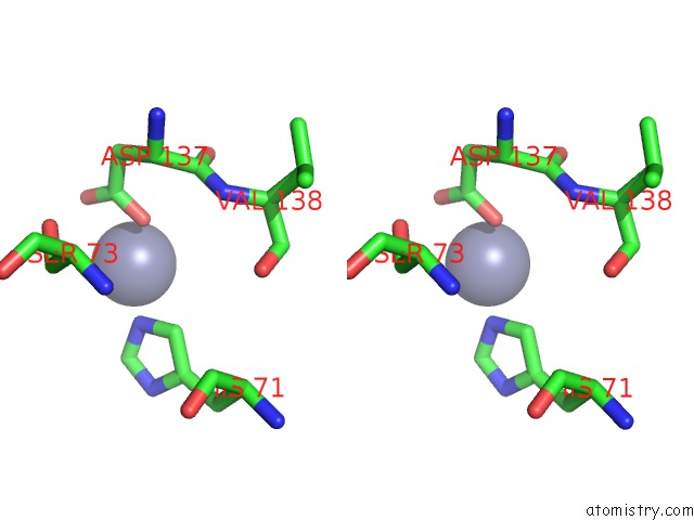
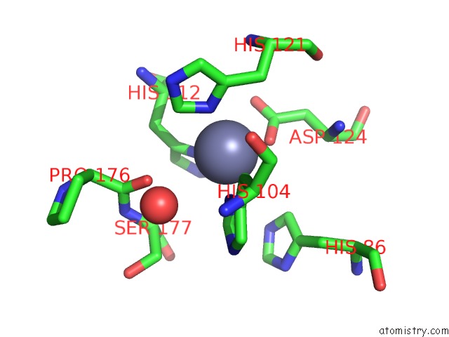
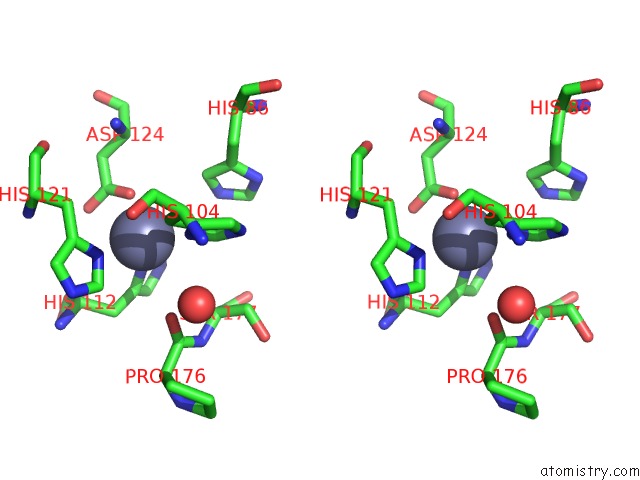
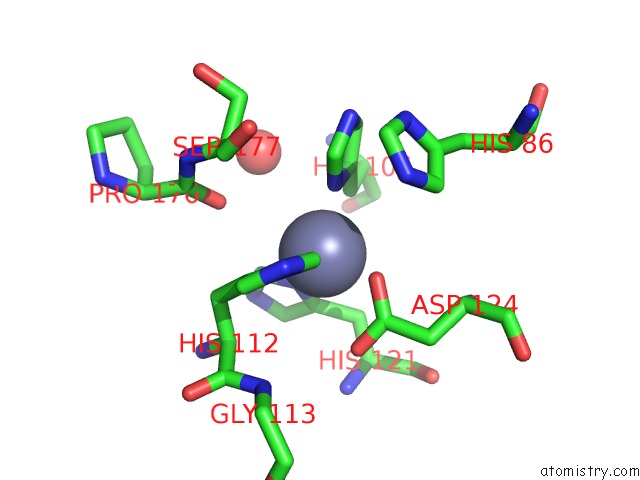
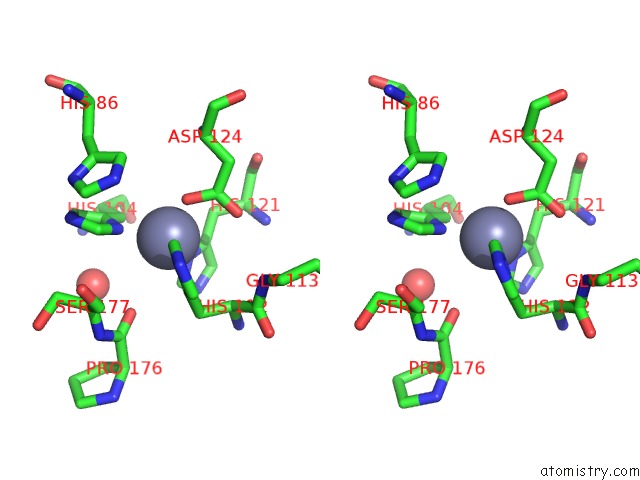
Zinc binding site 1 out of 10 in 1xtl
Go back to
Zinc binding site 1 out
of 10 in the Crystal Structure of P104H Mutant of Sod-Like Protein From Bacillus Subtilis.
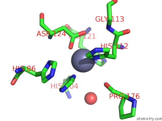
Mono view
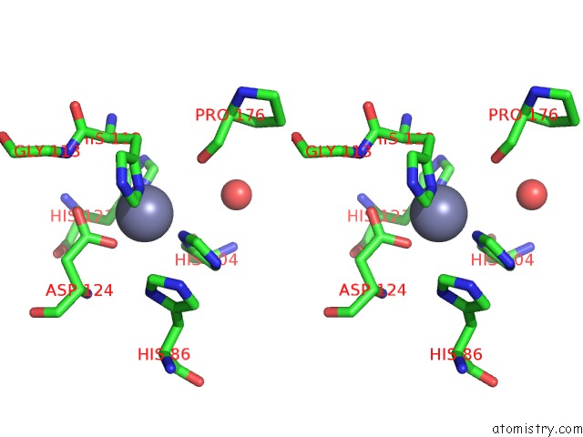
Stereo pair view
Mono view
Stereo pair view
A full contact list of Zinc with other atoms in the Zn binding
site number 1 of Crystal Structure of P104H Mutant of Sod-Like Protein From Bacillus Subtilis. within 5.0Å range:
|
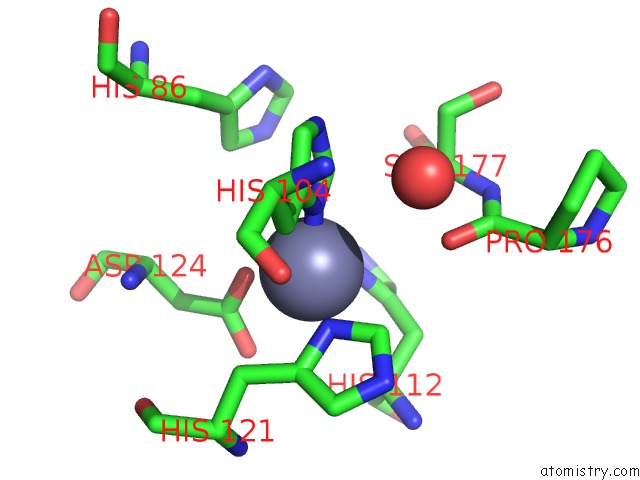
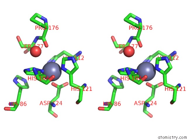
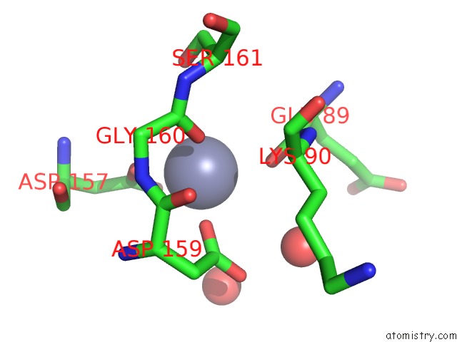
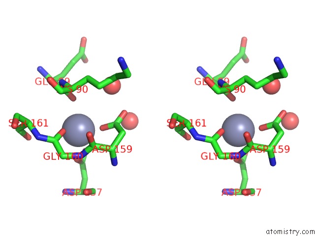
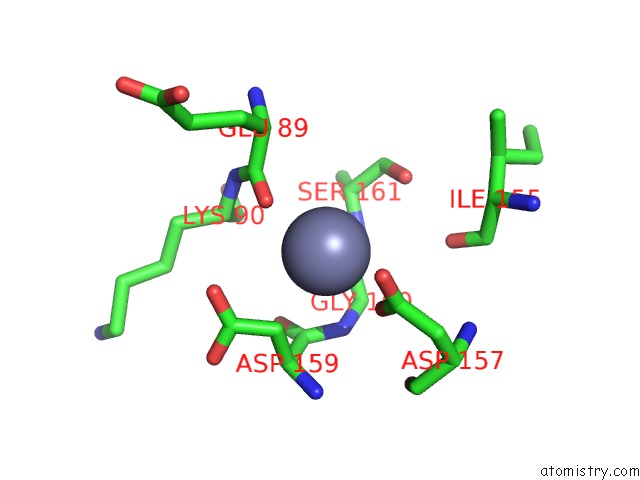
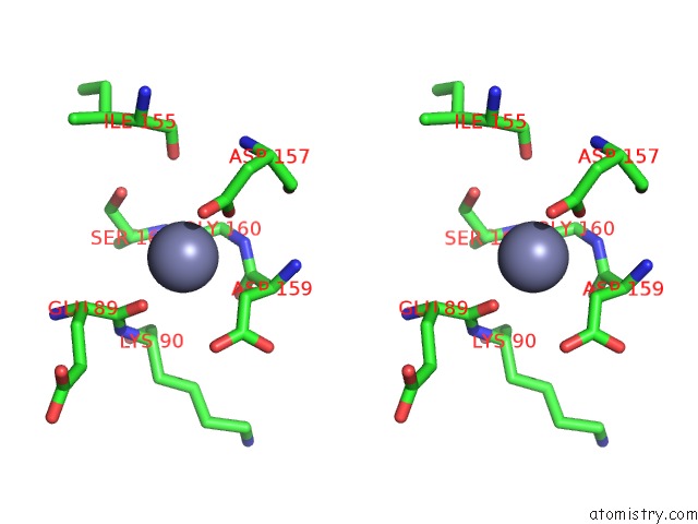
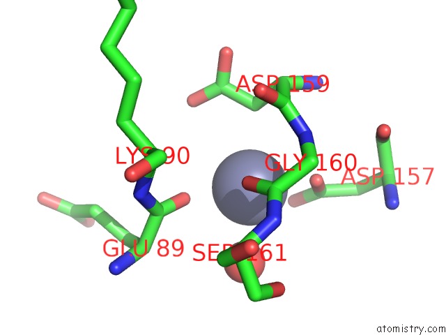
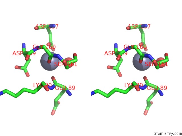
Zinc binding site 2 out of 10 in 1xtl
Go back to
Zinc binding site 2 out
of 10 in the Crystal Structure of P104H Mutant of Sod-Like Protein From Bacillus Subtilis.
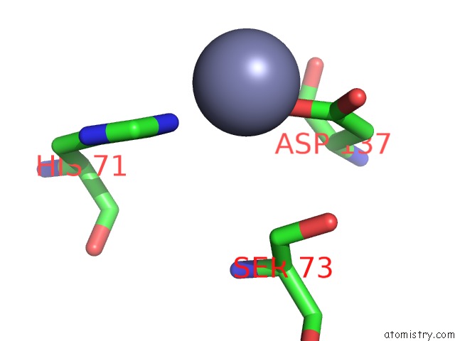
Mono view
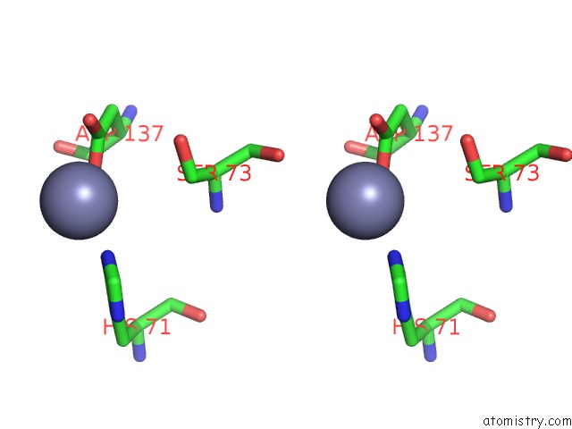
Stereo pair view
Mono view
Stereo pair view
A full contact list of Zinc with other atoms in the Zn binding
site number 2 of Crystal Structure of P104H Mutant of Sod-Like Protein From Bacillus Subtilis. within 5.0Å range:
|
Zinc binding site 3 out of 10 in 1xtl
Go back to
Zinc binding site 3 out
of 10 in the Crystal Structure of P104H Mutant of Sod-Like Protein From Bacillus Subtilis.
Mono view
Stereo pair view
Mono view
Stereo pair view
A full contact list of Zinc with other atoms in the Zn binding
site number 3 of Crystal Structure of P104H Mutant of Sod-Like Protein From Bacillus Subtilis. within 5.0Å range:
|
Zinc binding site 4 out of 10 in 1xtl
Go back to
Zinc binding site 4 out
of 10 in the Crystal Structure of P104H Mutant of Sod-Like Protein From Bacillus Subtilis.
Mono view
Stereo pair view
Mono view
Stereo pair view
A full contact list of Zinc with other atoms in the Zn binding
site number 4 of Crystal Structure of P104H Mutant of Sod-Like Protein From Bacillus Subtilis. within 5.0Å range:
|
Zinc binding site 5 out of 10 in 1xtl
Go back to
Zinc binding site 5 out
of 10 in the Crystal Structure of P104H Mutant of Sod-Like Protein From Bacillus Subtilis.
Mono view
Stereo pair view
Mono view
Stereo pair view
A full contact list of Zinc with other atoms in the Zn binding
site number 5 of Crystal Structure of P104H Mutant of Sod-Like Protein From Bacillus Subtilis. within 5.0Å range:
|
Zinc binding site 6 out of 10 in 1xtl
Go back to
Zinc binding site 6 out
of 10 in the Crystal Structure of P104H Mutant of Sod-Like Protein From Bacillus Subtilis.
Mono view
Stereo pair view
Mono view
Stereo pair view
A full contact list of Zinc with other atoms in the Zn binding
site number 6 of Crystal Structure of P104H Mutant of Sod-Like Protein From Bacillus Subtilis. within 5.0Å range:
|
Zinc binding site 7 out of 10 in 1xtl
Go back to
Zinc binding site 7 out
of 10 in the Crystal Structure of P104H Mutant of Sod-Like Protein From Bacillus Subtilis.
Mono view
Stereo pair view
Mono view
Stereo pair view
A full contact list of Zinc with other atoms in the Zn binding
site number 7 of Crystal Structure of P104H Mutant of Sod-Like Protein From Bacillus Subtilis. within 5.0Å range:
|
Zinc binding site 8 out of 10 in 1xtl
Go back to
Zinc binding site 8 out
of 10 in the Crystal Structure of P104H Mutant of Sod-Like Protein From Bacillus Subtilis.
Mono view
Stereo pair view
Mono view
Stereo pair view
A full contact list of Zinc with other atoms in the Zn binding
site number 8 of Crystal Structure of P104H Mutant of Sod-Like Protein From Bacillus Subtilis. within 5.0Å range:
|
Zinc binding site 9 out of 10 in 1xtl
Go back to
Zinc binding site 9 out
of 10 in the Crystal Structure of P104H Mutant of Sod-Like Protein From Bacillus Subtilis.
Mono view
Stereo pair view
Mono view
Stereo pair view
A full contact list of Zinc with other atoms in the Zn binding
site number 9 of Crystal Structure of P104H Mutant of Sod-Like Protein From Bacillus Subtilis. within 5.0Å range:
|
Zinc binding site 10 out of 10 in 1xtl
Go back to
Zinc binding site 10 out
of 10 in the Crystal Structure of P104H Mutant of Sod-Like Protein From Bacillus Subtilis.
Mono view
Stereo pair view
Mono view
Stereo pair view
A full contact list of Zinc with other atoms in the Zn binding
site number 10 of Crystal Structure of P104H Mutant of Sod-Like Protein From Bacillus Subtilis. within 5.0Å range:
|
Reference:
L.Banci,
M.Benvenuti,
I.Bertini,
D.E.Cabelli,
V.Calderone,
A.Fantoni,
S.Mangani,
M.Migliardi,
M.S.Viezzoli.
From An Inactive Prokaryotic Sod Homologue to An Active Protein Through Site-Directed Mutagenesis. J.Am.Chem.Soc. V. 127 13287 2005.
ISSN: ISSN 0002-7863
PubMed: 16173759
DOI: 10.1021/JA052790O
Page generated: Wed Oct 16 20:35:44 2024
ISSN: ISSN 0002-7863
PubMed: 16173759
DOI: 10.1021/JA052790O
Last articles
Zn in 9MJ5Zn in 9HNW
Zn in 9G0L
Zn in 9FNE
Zn in 9DZN
Zn in 9E0I
Zn in 9D32
Zn in 9DAK
Zn in 8ZXC
Zn in 8ZUF