Zinc in PDB 1wyh: Solution Structure of the Lim Domain From Human Skeletal Muscle Lim-Protein 2
Zinc Binding Sites:
The binding sites of Zinc atom in the Solution Structure of the Lim Domain From Human Skeletal Muscle Lim-Protein 2
(pdb code 1wyh). This binding sites where shown within
5.0 Angstroms radius around Zinc atom.
In total 2 binding sites of Zinc where determined in the
Solution Structure of the Lim Domain From Human Skeletal Muscle Lim-Protein 2, PDB code: 1wyh:
Jump to Zinc binding site number:
1;
2;
Zinc binding site 1 out
of 2 in 1wyh
Go back to
Zinc Binding Sites List in 1wyh
Zinc binding site 1 out
of 2 in the Solution Structure of the Lim Domain From Human Skeletal Muscle Lim-Protein 2
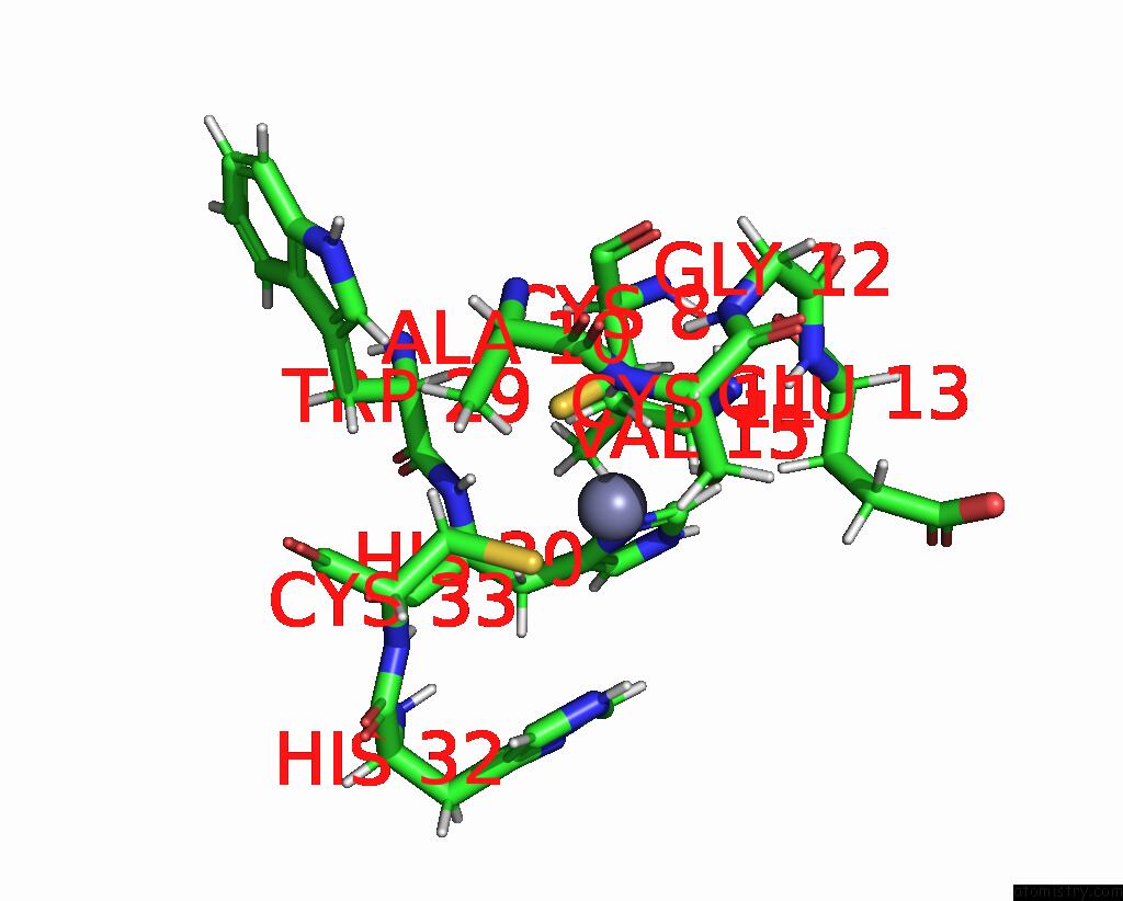
Mono view
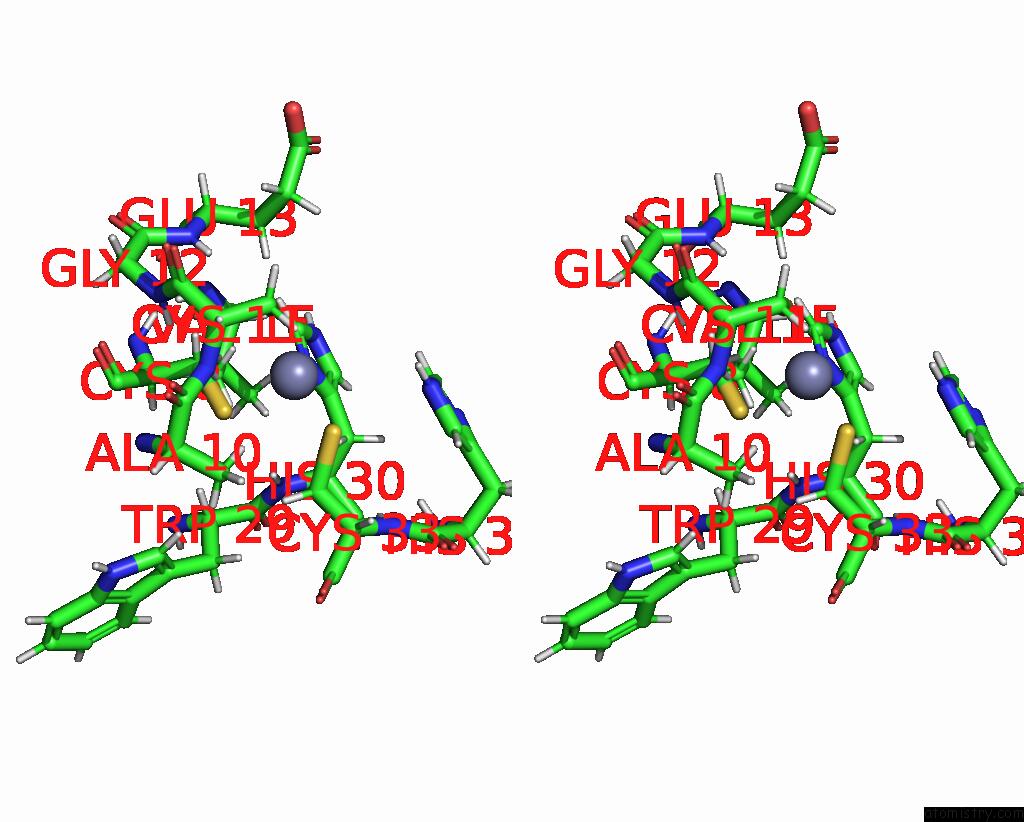
Stereo pair view
|
A full contact list of Zinc with other atoms in the Zn binding
site number 1 of Solution Structure of the Lim Domain From Human Skeletal Muscle Lim-Protein 2 within 5.0Å range:
probe
|
atom
|
residue
|
distance (Å)
|
B
|
Occ
|
A:Zn201
b:0.0
occ:1.00
|
SG
|
A:CYS33
|
2.3
|
0.0
|
1.0
|
SG
|
A:CYS11
|
2.3
|
0.0
|
1.0
|
ND1
|
A:HIS30
|
2.3
|
0.0
|
1.0
|
SG
|
A:CYS8
|
2.4
|
0.0
|
1.0
|
HB2
|
A:HIS30
|
2.8
|
0.0
|
1.0
|
H
|
A:CYS11
|
3.0
|
0.0
|
1.0
|
CB
|
A:CYS8
|
3.2
|
0.0
|
1.0
|
HB3
|
A:CYS8
|
3.2
|
0.0
|
1.0
|
HB2
|
A:CYS8
|
3.3
|
0.0
|
1.0
|
CE1
|
A:HIS30
|
3.3
|
0.0
|
1.0
|
CG
|
A:HIS30
|
3.3
|
0.0
|
1.0
|
HB3
|
A:ALA10
|
3.3
|
0.0
|
1.0
|
HB2
|
A:CYS33
|
3.4
|
0.0
|
1.0
|
HB3
|
A:CYS11
|
3.5
|
0.0
|
1.0
|
HE1
|
A:HIS30
|
3.5
|
0.0
|
1.0
|
CB
|
A:CYS33
|
3.5
|
0.0
|
1.0
|
CB
|
A:CYS11
|
3.5
|
0.0
|
1.0
|
CB
|
A:HIS30
|
3.6
|
0.0
|
1.0
|
H
|
A:HIS30
|
3.6
|
0.0
|
1.0
|
HE2
|
A:HIS32
|
3.7
|
0.0
|
1.0
|
HB3
|
A:CYS33
|
3.8
|
0.0
|
1.0
|
N
|
A:CYS11
|
3.8
|
0.0
|
1.0
|
HB2
|
A:GLU13
|
3.8
|
0.0
|
1.0
|
NE2
|
A:HIS32
|
4.0
|
0.0
|
1.0
|
H
|
A:GLY12
|
4.1
|
0.0
|
1.0
|
HE1
|
A:HIS32
|
4.1
|
0.0
|
1.0
|
HB3
|
A:HIS30
|
4.2
|
0.0
|
1.0
|
CE1
|
A:HIS32
|
4.3
|
0.0
|
1.0
|
CA
|
A:CYS11
|
4.3
|
0.0
|
1.0
|
HB2
|
A:CYS11
|
4.4
|
0.0
|
1.0
|
N
|
A:HIS30
|
4.4
|
0.0
|
1.0
|
CB
|
A:ALA10
|
4.4
|
0.0
|
1.0
|
H
|
A:GLU13
|
4.4
|
0.0
|
1.0
|
NE2
|
A:HIS30
|
4.4
|
0.0
|
1.0
|
H
|
A:ALA10
|
4.4
|
0.0
|
1.0
|
CD2
|
A:HIS30
|
4.4
|
0.0
|
1.0
|
CA
|
A:HIS30
|
4.6
|
0.0
|
1.0
|
CA
|
A:CYS8
|
4.7
|
0.0
|
1.0
|
CA
|
A:CYS33
|
4.8
|
0.0
|
1.0
|
N
|
A:GLY12
|
4.8
|
0.0
|
1.0
|
HG3
|
A:GLU13
|
4.8
|
0.0
|
1.0
|
HG22
|
A:VAL15
|
4.9
|
0.0
|
1.0
|
HB2
|
A:ALA10
|
4.9
|
0.0
|
1.0
|
C
|
A:ALA10
|
4.9
|
0.0
|
1.0
|
HB1
|
A:ALA10
|
4.9
|
0.0
|
1.0
|
CB
|
A:GLU13
|
4.9
|
0.0
|
1.0
|
CD2
|
A:HIS32
|
4.9
|
0.0
|
1.0
|
HA
|
A:TRP29
|
5.0
|
0.0
|
1.0
|
H
|
A:CYS33
|
5.0
|
0.0
|
1.0
|
|
Zinc binding site 2 out
of 2 in 1wyh
Go back to
Zinc Binding Sites List in 1wyh
Zinc binding site 2 out
of 2 in the Solution Structure of the Lim Domain From Human Skeletal Muscle Lim-Protein 2
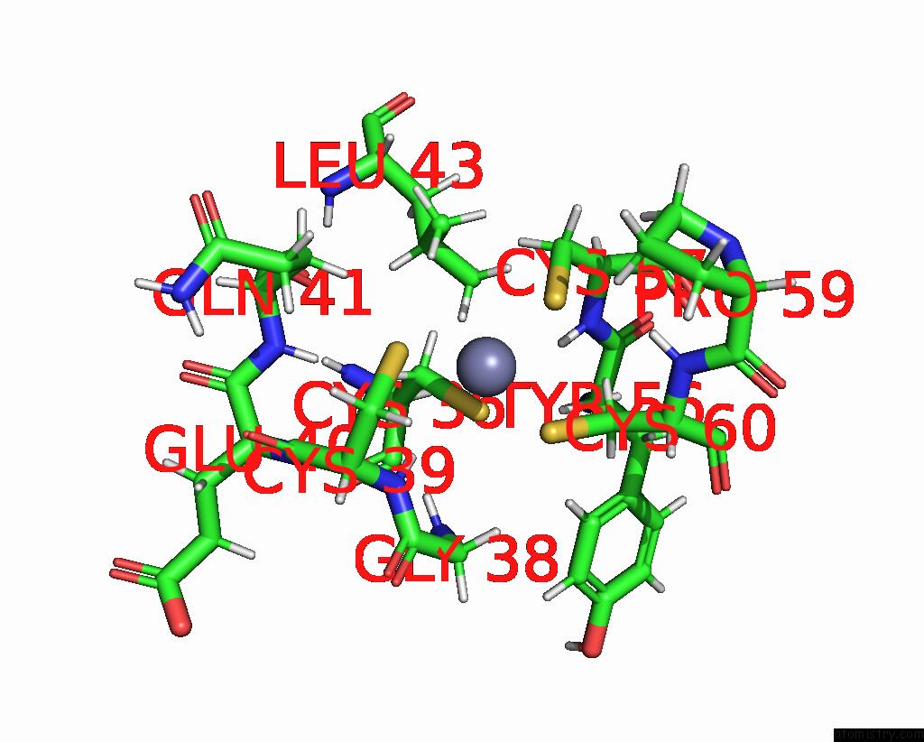
Mono view
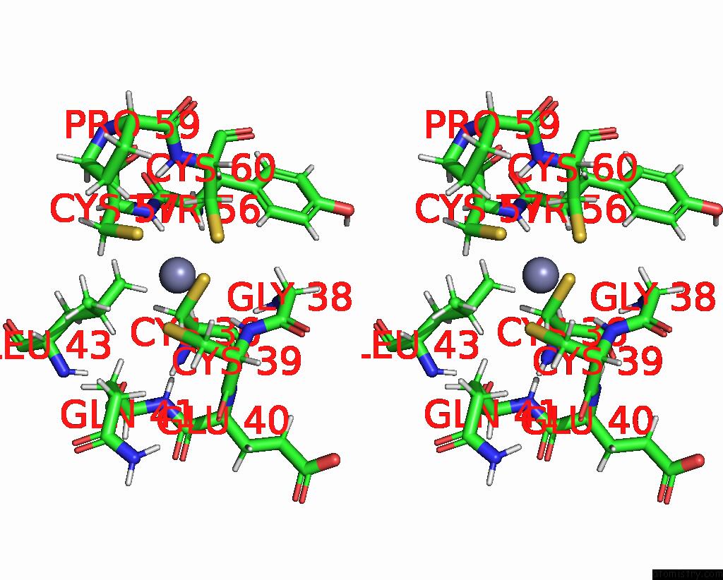
Stereo pair view
|
A full contact list of Zinc with other atoms in the Zn binding
site number 2 of Solution Structure of the Lim Domain From Human Skeletal Muscle Lim-Protein 2 within 5.0Å range:
probe
|
atom
|
residue
|
distance (Å)
|
B
|
Occ
|
A:Zn401
b:0.0
occ:1.00
|
SG
|
A:CYS57
|
2.3
|
0.0
|
1.0
|
SG
|
A:CYS39
|
2.3
|
0.0
|
1.0
|
SG
|
A:CYS60
|
2.3
|
0.0
|
1.0
|
SG
|
A:CYS36
|
2.4
|
0.0
|
1.0
|
H
|
A:CYS39
|
2.8
|
0.0
|
1.0
|
H
|
A:CYS57
|
2.9
|
0.0
|
1.0
|
HB2
|
A:CYS60
|
3.0
|
0.0
|
1.0
|
HB3
|
A:CYS36
|
3.1
|
0.0
|
1.0
|
CB
|
A:CYS36
|
3.2
|
0.0
|
1.0
|
CB
|
A:CYS60
|
3.2
|
0.0
|
1.0
|
HB2
|
A:CYS36
|
3.3
|
0.0
|
1.0
|
HB3
|
A:CYS57
|
3.5
|
0.0
|
1.0
|
HB3
|
A:CYS39
|
3.5
|
0.0
|
1.0
|
CB
|
A:CYS39
|
3.5
|
0.0
|
1.0
|
N
|
A:CYS39
|
3.5
|
0.0
|
1.0
|
CB
|
A:CYS57
|
3.6
|
0.0
|
1.0
|
HB3
|
A:CYS60
|
3.8
|
0.0
|
1.0
|
N
|
A:CYS57
|
3.8
|
0.0
|
1.0
|
HA3
|
A:GLY38
|
3.9
|
0.0
|
1.0
|
H
|
A:GLY38
|
4.1
|
0.0
|
1.0
|
HB2
|
A:GLN41
|
4.1
|
0.0
|
1.0
|
H
|
A:CYS60
|
4.1
|
0.0
|
1.0
|
HD22
|
A:LEU43
|
4.1
|
0.0
|
1.0
|
CA
|
A:CYS39
|
4.1
|
0.0
|
1.0
|
HA
|
A:TYR56
|
4.2
|
0.0
|
1.0
|
HG2
|
A:PRO59
|
4.2
|
0.0
|
1.0
|
CA
|
A:CYS57
|
4.3
|
0.0
|
1.0
|
H
|
A:GLN41
|
4.4
|
0.0
|
1.0
|
HB2
|
A:CYS57
|
4.4
|
0.0
|
1.0
|
HB2
|
A:CYS39
|
4.4
|
0.0
|
1.0
|
C
|
A:GLY38
|
4.4
|
0.0
|
1.0
|
H
|
A:GLU40
|
4.5
|
0.0
|
1.0
|
CA
|
A:GLY38
|
4.5
|
0.0
|
1.0
|
CA
|
A:CYS60
|
4.5
|
0.0
|
1.0
|
HD1
|
A:TYR56
|
4.6
|
0.0
|
1.0
|
N
|
A:GLY38
|
4.6
|
0.0
|
1.0
|
CA
|
A:CYS36
|
4.7
|
0.0
|
1.0
|
N
|
A:CYS60
|
4.7
|
0.0
|
1.0
|
HA
|
A:CYS60
|
4.9
|
0.0
|
1.0
|
C
|
A:TYR56
|
4.9
|
0.0
|
1.0
|
C
|
A:CYS39
|
4.9
|
0.0
|
1.0
|
HG2
|
A:GLN41
|
5.0
|
0.0
|
1.0
|
N
|
A:GLU40
|
5.0
|
0.0
|
1.0
|
HA
|
A:CYS39
|
5.0
|
0.0
|
1.0
|
|
Reference:
T.N.Niraula,
N.Tochio,
S.Koshiba,
M.Inoue,
T.Kigawa,
S.Yokoyama.
Solution Structure of the Lim Domain From Human Skeletal Muscle Lim-Protein 2 To Be Published.
Page generated: Wed Oct 16 20:13:10 2024
|