Zinc »
PDB 1wwe-1x8g »
1wxo »
Zinc in PDB 1wxo: Structure of Archaeal Trans-Editing Protein Alax in Complex with Zinc
Protein crystallography data
The structure of Structure of Archaeal Trans-Editing Protein Alax in Complex with Zinc, PDB code: 1wxo
was solved by
M.Sokabe,
A.Okada,
T.Nakashima,
M.Yao,
I.Tanaka,
with X-Ray Crystallography technique. A brief refinement statistics is given in the table below:
| Resolution Low / High (Å) | 50.00 / 1.88 |
| Space group | P 1 21 1 |
| Cell size a, b, c (Å), α, β, γ (°) | 42.472, 97.971, 60.584, 90.00, 95.81, 90.00 |
| R / Rfree (%) | 19.3 / 23.2 |
Zinc Binding Sites:
The binding sites of Zinc atom in the Structure of Archaeal Trans-Editing Protein Alax in Complex with Zinc
(pdb code 1wxo). This binding sites where shown within
5.0 Angstroms radius around Zinc atom.
In total 3 binding sites of Zinc where determined in the Structure of Archaeal Trans-Editing Protein Alax in Complex with Zinc, PDB code: 1wxo:
Jump to Zinc binding site number: 1; 2; 3;
In total 3 binding sites of Zinc where determined in the Structure of Archaeal Trans-Editing Protein Alax in Complex with Zinc, PDB code: 1wxo:
Jump to Zinc binding site number: 1; 2; 3;
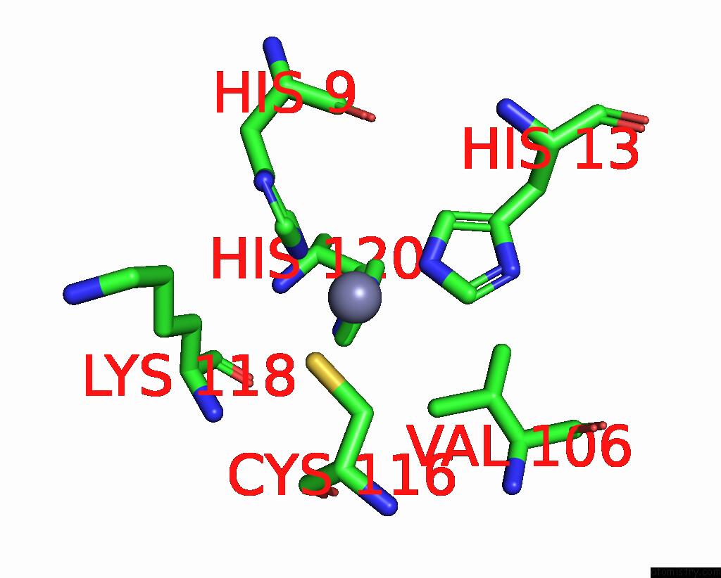
Zinc binding site 1 out of 3 in 1wxo
Go back to
Zinc binding site 1 out
of 3 in the Structure of Archaeal Trans-Editing Protein Alax in Complex with Zinc
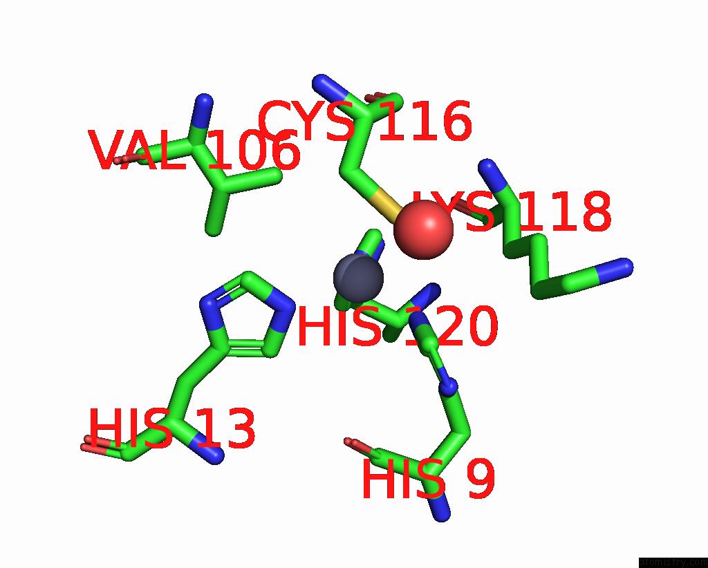
Mono view
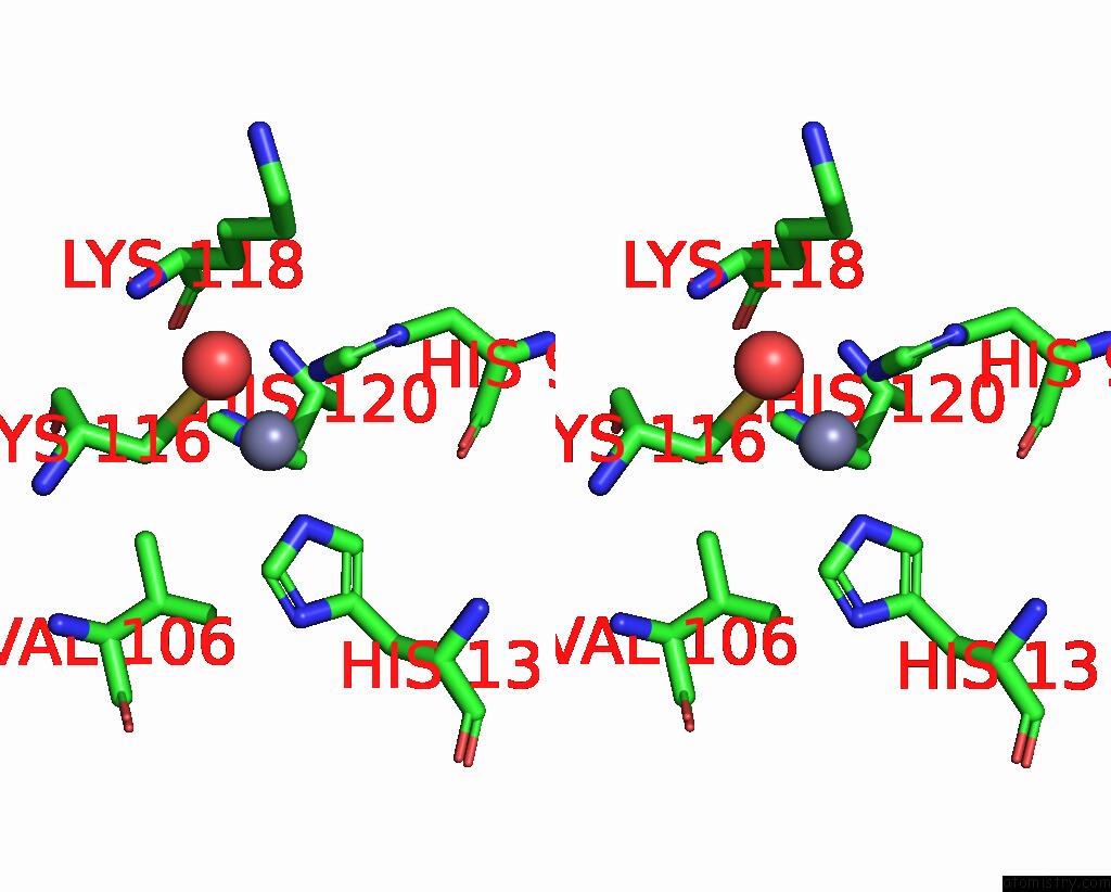
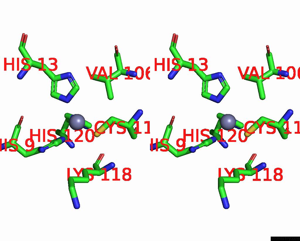
Stereo pair view
Mono view
Stereo pair view
A full contact list of Zinc with other atoms in the Zn binding
site number 1 of Structure of Archaeal Trans-Editing Protein Alax in Complex with Zinc within 5.0Å range:
|
Zinc binding site 2 out of 3 in 1wxo
Go back to
Zinc binding site 2 out
of 3 in the Structure of Archaeal Trans-Editing Protein Alax in Complex with Zinc
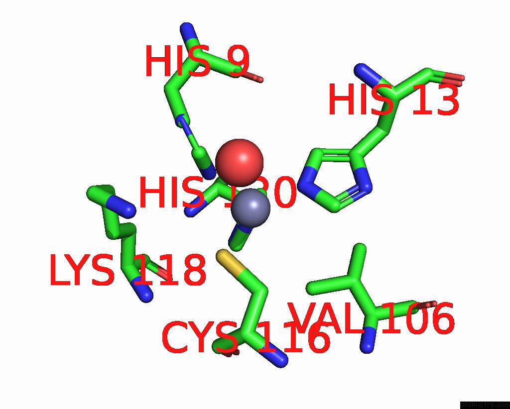
Mono view
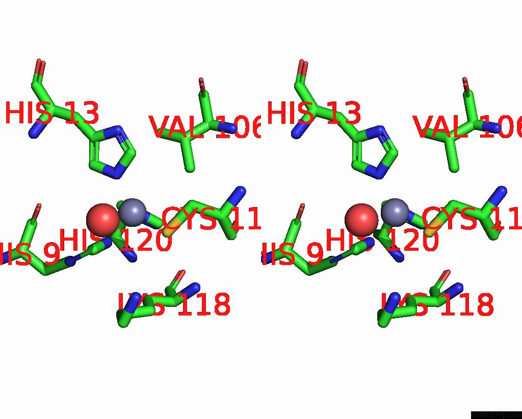
Stereo pair view
Mono view
Stereo pair view
A full contact list of Zinc with other atoms in the Zn binding
site number 2 of Structure of Archaeal Trans-Editing Protein Alax in Complex with Zinc within 5.0Å range:
|
Zinc binding site 3 out of 3 in 1wxo
Go back to
Zinc binding site 3 out
of 3 in the Structure of Archaeal Trans-Editing Protein Alax in Complex with Zinc
Mono view
Stereo pair view
Mono view
Stereo pair view
A full contact list of Zinc with other atoms in the Zn binding
site number 3 of Structure of Archaeal Trans-Editing Protein Alax in Complex with Zinc within 5.0Å range:
|
Reference:
M.Sokabe,
A.Okada,
M.Yao,
T.Nakashima,
I.Tanaka.
Molecular Basis of Alanine Discrimination in Editing Site Proc.Natl.Acad.Sci.Usa V. 102 11669 2005.
ISSN: ISSN 0027-8424
PubMed: 16087889
DOI: 10.1073/PNAS.0502119102
Page generated: Wed Aug 20 00:08:05 2025
ISSN: ISSN 0027-8424
PubMed: 16087889
DOI: 10.1073/PNAS.0502119102
Last articles
Zn in 2Z29Zn in 2Z28
Zn in 2Z2B
Zn in 2Z27
Zn in 2Z26
Zn in 2Z24
Zn in 2Z25
Zn in 2Z0P
Zn in 2Z1X
Zn in 2Z1A