Zinc »
PDB 1w5m-1wfx »
1waa »
Zinc in PDB 1waa: IG27 Protein Domain
Protein crystallography data
The structure of IG27 Protein Domain, PDB code: 1waa
was solved by
M.C.Vega,
L.Valencia,
P.Zou,
M.Wilmanns,
with X-Ray Crystallography technique. A brief refinement statistics is given in the table below:
| Resolution Low / High (Å) | 15.00 / 1.80 |
| Space group | P 21 21 21 |
| Cell size a, b, c (Å), α, β, γ (°) | 62.240, 75.990, 134.220, 90.00, 90.00, 90.00 |
| R / Rfree (%) | 20.7 / 26.8 |
Zinc Binding Sites:
Pages:
>>> Page 1 <<< Page 2, Binding sites: 11 - 20; Page 3, Binding sites: 21 - 24;Binding sites:
The binding sites of Zinc atom in the IG27 Protein Domain (pdb code 1waa). This binding sites where shown within 5.0 Angstroms radius around Zinc atom.In total 24 binding sites of Zinc where determined in the IG27 Protein Domain, PDB code: 1waa:
Jump to Zinc binding site number: 1; 2; 3; 4; 5; 6; 7; 8; 9; 10;
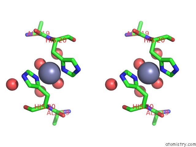
Zinc binding site 1 out of 24 in 1waa
Go back to
Zinc binding site 1 out
of 24 in the IG27 Protein Domain
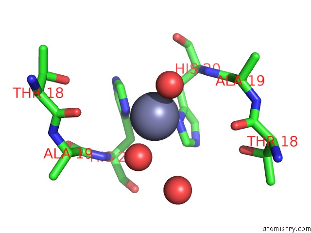
Mono view
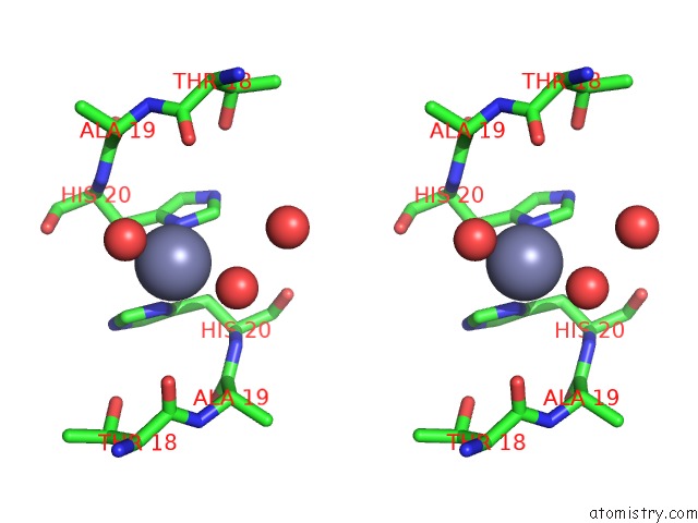
Stereo pair view
Mono view
Stereo pair view
A full contact list of Zinc with other atoms in the Zn binding
site number 1 of IG27 Protein Domain within 5.0Å range:
|
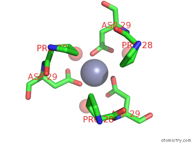
Zinc binding site 2 out of 24 in 1waa
Go back to
Zinc binding site 2 out
of 24 in the IG27 Protein Domain

Mono view

Stereo pair view
Mono view
Stereo pair view
A full contact list of Zinc with other atoms in the Zn binding
site number 2 of IG27 Protein Domain within 5.0Å range:
|
Zinc binding site 3 out of 24 in 1waa
Go back to
Zinc binding site 3 out
of 24 in the IG27 Protein Domain
Mono view

Stereo pair view
Mono view
Stereo pair view
A full contact list of Zinc with other atoms in the Zn binding
site number 3 of IG27 Protein Domain within 5.0Å range:
|
Zinc binding site 4 out of 24 in 1waa
Go back to
Zinc binding site 4 out
of 24 in the IG27 Protein Domain

Mono view

Stereo pair view
Mono view
Stereo pair view
A full contact list of Zinc with other atoms in the Zn binding
site number 4 of IG27 Protein Domain within 5.0Å range:
|
Zinc binding site 5 out of 24 in 1waa
Go back to
Zinc binding site 5 out
of 24 in the IG27 Protein Domain

Mono view
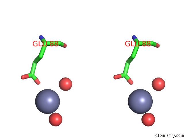
Stereo pair view
Mono view
Stereo pair view
A full contact list of Zinc with other atoms in the Zn binding
site number 5 of IG27 Protein Domain within 5.0Å range:
|
Zinc binding site 6 out of 24 in 1waa
Go back to
Zinc binding site 6 out
of 24 in the IG27 Protein Domain

Mono view

Stereo pair view
Mono view
Stereo pair view
A full contact list of Zinc with other atoms in the Zn binding
site number 6 of IG27 Protein Domain within 5.0Å range:
|
Zinc binding site 7 out of 24 in 1waa
Go back to
Zinc binding site 7 out
of 24 in the IG27 Protein Domain

Mono view

Stereo pair view
Mono view
Stereo pair view
A full contact list of Zinc with other atoms in the Zn binding
site number 7 of IG27 Protein Domain within 5.0Å range:
|
Zinc binding site 8 out of 24 in 1waa
Go back to
Zinc binding site 8 out
of 24 in the IG27 Protein Domain

Mono view

Stereo pair view
Mono view
Stereo pair view
A full contact list of Zinc with other atoms in the Zn binding
site number 8 of IG27 Protein Domain within 5.0Å range:
|
Zinc binding site 9 out of 24 in 1waa
Go back to
Zinc binding site 9 out
of 24 in the IG27 Protein Domain

Mono view
Stereo pair view
Mono view
Stereo pair view
A full contact list of Zinc with other atoms in the Zn binding
site number 9 of IG27 Protein Domain within 5.0Å range:
|
Zinc binding site 10 out of 24 in 1waa
Go back to
Zinc binding site 10 out
of 24 in the IG27 Protein Domain

Mono view

Stereo pair view
Mono view
Stereo pair view
A full contact list of Zinc with other atoms in the Zn binding
site number 10 of IG27 Protein Domain within 5.0Å range:
|
Reference:
W.Stacklies,
M.C.Vega,
M.Wilmanns,
F.Grater.
Mechanical Network in Titin Immunoglobulin From Force Distribution Analysis. Plos Comput.Biol. V. 5 00306 2009.
ISSN: ISSN 1553-734X
PubMed: 19282960
DOI: 10.1371/JOURNAL.PCBI.1000306
Page generated: Wed Oct 16 19:57:10 2024
ISSN: ISSN 1553-734X
PubMed: 19282960
DOI: 10.1371/JOURNAL.PCBI.1000306
Last articles
Zn in 9MJ5Zn in 9HNW
Zn in 9G0L
Zn in 9FNE
Zn in 9DZN
Zn in 9E0I
Zn in 9D32
Zn in 9DAK
Zn in 8ZXC
Zn in 8ZUF