Zinc »
PDB 1vev-1w57 »
1w1i »
Zinc in PDB 1w1i: Crystal Structure of Dipeptidyl Peptidase IV (Dppiv or CD26) in Complex with Adenosine Deaminase
Enzymatic activity of Crystal Structure of Dipeptidyl Peptidase IV (Dppiv or CD26) in Complex with Adenosine Deaminase
All present enzymatic activity of Crystal Structure of Dipeptidyl Peptidase IV (Dppiv or CD26) in Complex with Adenosine Deaminase:
3.4.14.5; 3.5.4.4;
3.4.14.5; 3.5.4.4;
Protein crystallography data
The structure of Crystal Structure of Dipeptidyl Peptidase IV (Dppiv or CD26) in Complex with Adenosine Deaminase, PDB code: 1w1i
was solved by
W.A.Weihofen,
J.Liu,
W.Reutter,
W.Saenger,
H.Fan,
with X-Ray Crystallography technique. A brief refinement statistics is given in the table below:
| Resolution Low / High (Å) | 30.00 / 3.03 |
| Space group | C 1 2 1 |
| Cell size a, b, c (Å), α, β, γ (°) | 158.065, 168.504, 236.842, 90.00, 100.54, 90.00 |
| R / Rfree (%) | 22.4 / 25.7 |
Zinc Binding Sites:
The binding sites of Zinc atom in the Crystal Structure of Dipeptidyl Peptidase IV (Dppiv or CD26) in Complex with Adenosine Deaminase
(pdb code 1w1i). This binding sites where shown within
5.0 Angstroms radius around Zinc atom.
In total 4 binding sites of Zinc where determined in the Crystal Structure of Dipeptidyl Peptidase IV (Dppiv or CD26) in Complex with Adenosine Deaminase, PDB code: 1w1i:
Jump to Zinc binding site number: 1; 2; 3; 4;
In total 4 binding sites of Zinc where determined in the Crystal Structure of Dipeptidyl Peptidase IV (Dppiv or CD26) in Complex with Adenosine Deaminase, PDB code: 1w1i:
Jump to Zinc binding site number: 1; 2; 3; 4;
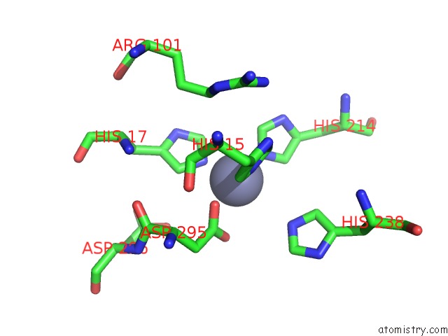
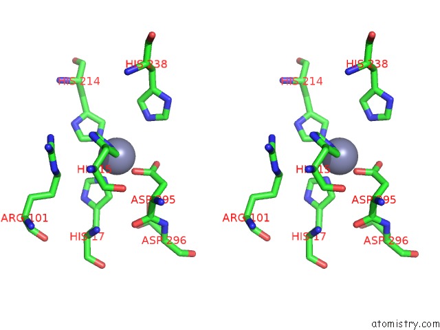
Zinc binding site 1 out of 4 in 1w1i
Go back to
Zinc binding site 1 out
of 4 in the Crystal Structure of Dipeptidyl Peptidase IV (Dppiv or CD26) in Complex with Adenosine Deaminase
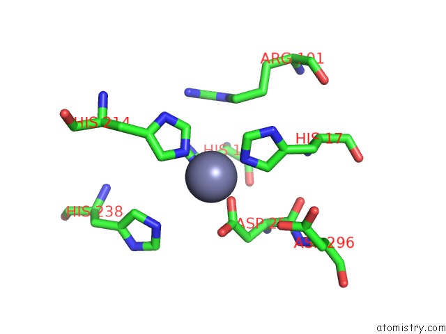
Mono view
Stereo pair view
Mono view
Stereo pair view
A full contact list of Zinc with other atoms in the Zn binding
site number 1 of Crystal Structure of Dipeptidyl Peptidase IV (Dppiv or CD26) in Complex with Adenosine Deaminase within 5.0Å range:
|
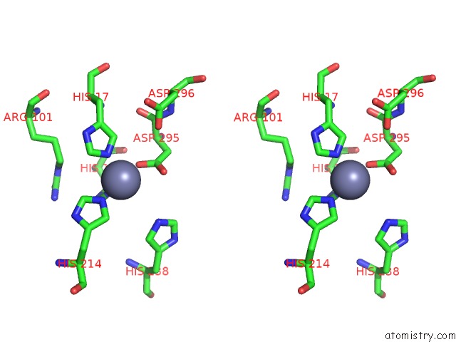
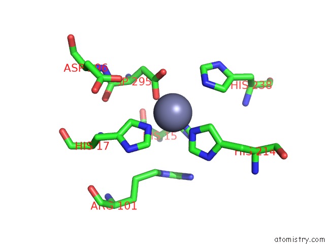
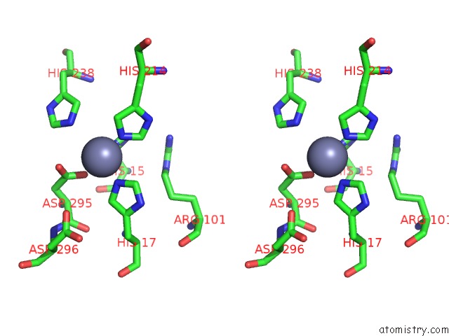
Zinc binding site 2 out of 4 in 1w1i
Go back to
Zinc binding site 2 out
of 4 in the Crystal Structure of Dipeptidyl Peptidase IV (Dppiv or CD26) in Complex with Adenosine Deaminase
Mono view
Stereo pair view
Mono view
Stereo pair view
A full contact list of Zinc with other atoms in the Zn binding
site number 2 of Crystal Structure of Dipeptidyl Peptidase IV (Dppiv or CD26) in Complex with Adenosine Deaminase within 5.0Å range:
|
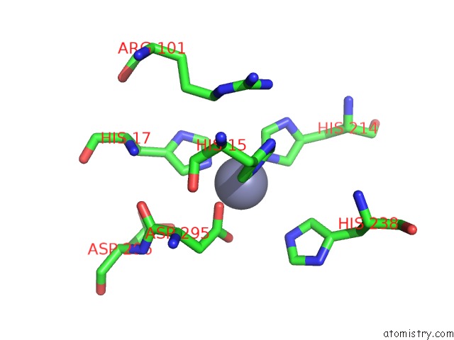
Zinc binding site 3 out of 4 in 1w1i
Go back to
Zinc binding site 3 out
of 4 in the Crystal Structure of Dipeptidyl Peptidase IV (Dppiv or CD26) in Complex with Adenosine Deaminase
Mono view
Stereo pair view
Mono view
Stereo pair view
A full contact list of Zinc with other atoms in the Zn binding
site number 3 of Crystal Structure of Dipeptidyl Peptidase IV (Dppiv or CD26) in Complex with Adenosine Deaminase within 5.0Å range:
|
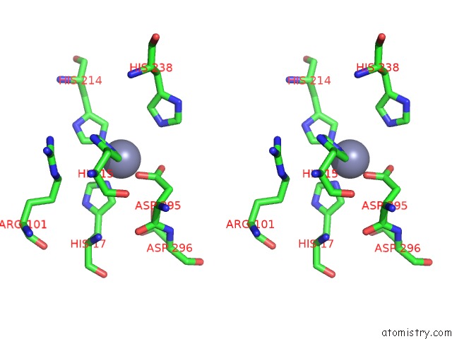
Zinc binding site 4 out of 4 in 1w1i
Go back to
Zinc binding site 4 out
of 4 in the Crystal Structure of Dipeptidyl Peptidase IV (Dppiv or CD26) in Complex with Adenosine Deaminase
Mono view
Stereo pair view
Mono view
Stereo pair view
A full contact list of Zinc with other atoms in the Zn binding
site number 4 of Crystal Structure of Dipeptidyl Peptidase IV (Dppiv or CD26) in Complex with Adenosine Deaminase within 5.0Å range:
|
Reference:
W.A.Weihofen,
J.Liu,
W.Reutter,
W.Saenger,
H.Fan.
Crystal Structure of CD26/Dipeptidyl-Peptidase IV in Complex with Adenosine Deaminase Reveals A Highly Amphiphilic Interface. J. Biol. Chem. V. 279 43330 2004.
ISSN: ISSN 0021-9258
PubMed: 15213224
DOI: 10.1074/JBC.M405001200
Page generated: Wed Oct 16 19:53:42 2024
ISSN: ISSN 0021-9258
PubMed: 15213224
DOI: 10.1074/JBC.M405001200
Last articles
Zn in 9MJ5Zn in 9HNW
Zn in 9G0L
Zn in 9FNE
Zn in 9DZN
Zn in 9E0I
Zn in 9D32
Zn in 9DAK
Zn in 8ZXC
Zn in 8ZUF