Zinc »
PDB 1vev-1w57 »
1vs0 »
Zinc in PDB 1vs0: Crystal Structure of the Ligase Domain From M. Tuberculosis Ligd at 2.4A
Protein crystallography data
The structure of Crystal Structure of the Ligase Domain From M. Tuberculosis Ligd at 2.4A, PDB code: 1vs0
was solved by
D.Akey,
A.Martins,
J.Aniukwu,
M.S.Glickman,
S.Shuman,
J.M.Berger,
Tbstructural Genomics Consortium (Tbsgc),
with X-Ray Crystallography technique. A brief refinement statistics is given in the table below:
| Resolution Low / High (Å) | 50.00 / 2.40 |
| Space group | P 32 2 1 |
| Cell size a, b, c (Å), α, β, γ (°) | 57.102, 57.102, 368.957, 90.00, 90.00, 120.00 |
| R / Rfree (%) | 19.2 / 24.8 |
Other elements in 1vs0:
The structure of Crystal Structure of the Ligase Domain From M. Tuberculosis Ligd at 2.4A also contains other interesting chemical elements:
| Magnesium | (Mg) | 1 atom |
| Chlorine | (Cl) | 4 atoms |
Zinc Binding Sites:
The binding sites of Zinc atom in the Crystal Structure of the Ligase Domain From M. Tuberculosis Ligd at 2.4A
(pdb code 1vs0). This binding sites where shown within
5.0 Angstroms radius around Zinc atom.
In total 4 binding sites of Zinc where determined in the Crystal Structure of the Ligase Domain From M. Tuberculosis Ligd at 2.4A, PDB code: 1vs0:
Jump to Zinc binding site number: 1; 2; 3; 4;
In total 4 binding sites of Zinc where determined in the Crystal Structure of the Ligase Domain From M. Tuberculosis Ligd at 2.4A, PDB code: 1vs0:
Jump to Zinc binding site number: 1; 2; 3; 4;
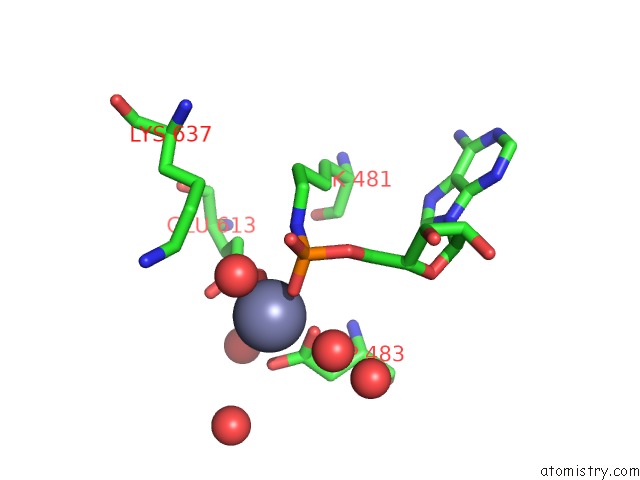
Zinc binding site 1 out of 4 in 1vs0
Go back to
Zinc binding site 1 out
of 4 in the Crystal Structure of the Ligase Domain From M. Tuberculosis Ligd at 2.4A
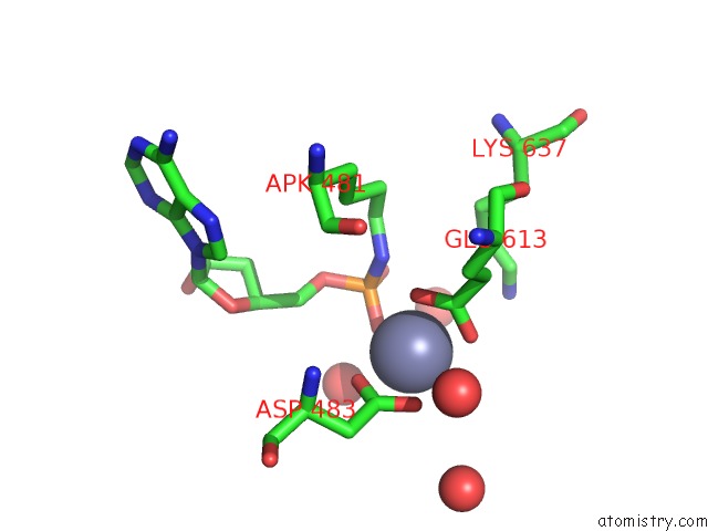
Mono view
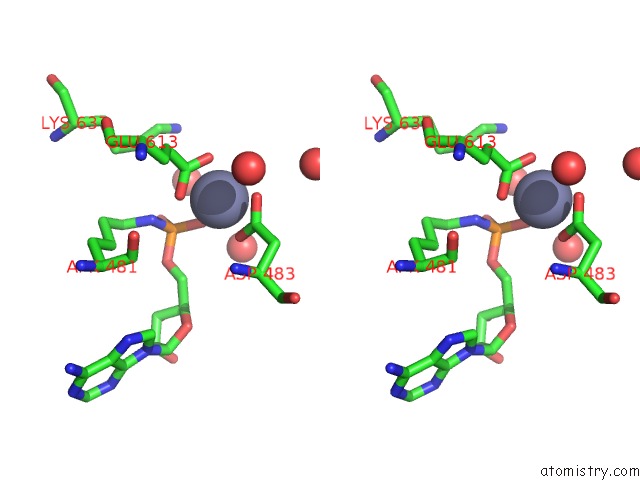
Stereo pair view
Mono view
Stereo pair view
A full contact list of Zinc with other atoms in the Zn binding
site number 1 of Crystal Structure of the Ligase Domain From M. Tuberculosis Ligd at 2.4A within 5.0Å range:
|
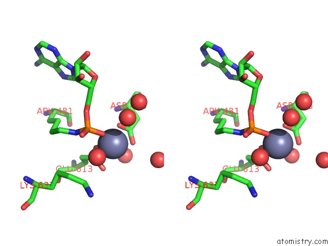
Zinc binding site 2 out of 4 in 1vs0
Go back to
Zinc binding site 2 out
of 4 in the Crystal Structure of the Ligase Domain From M. Tuberculosis Ligd at 2.4A
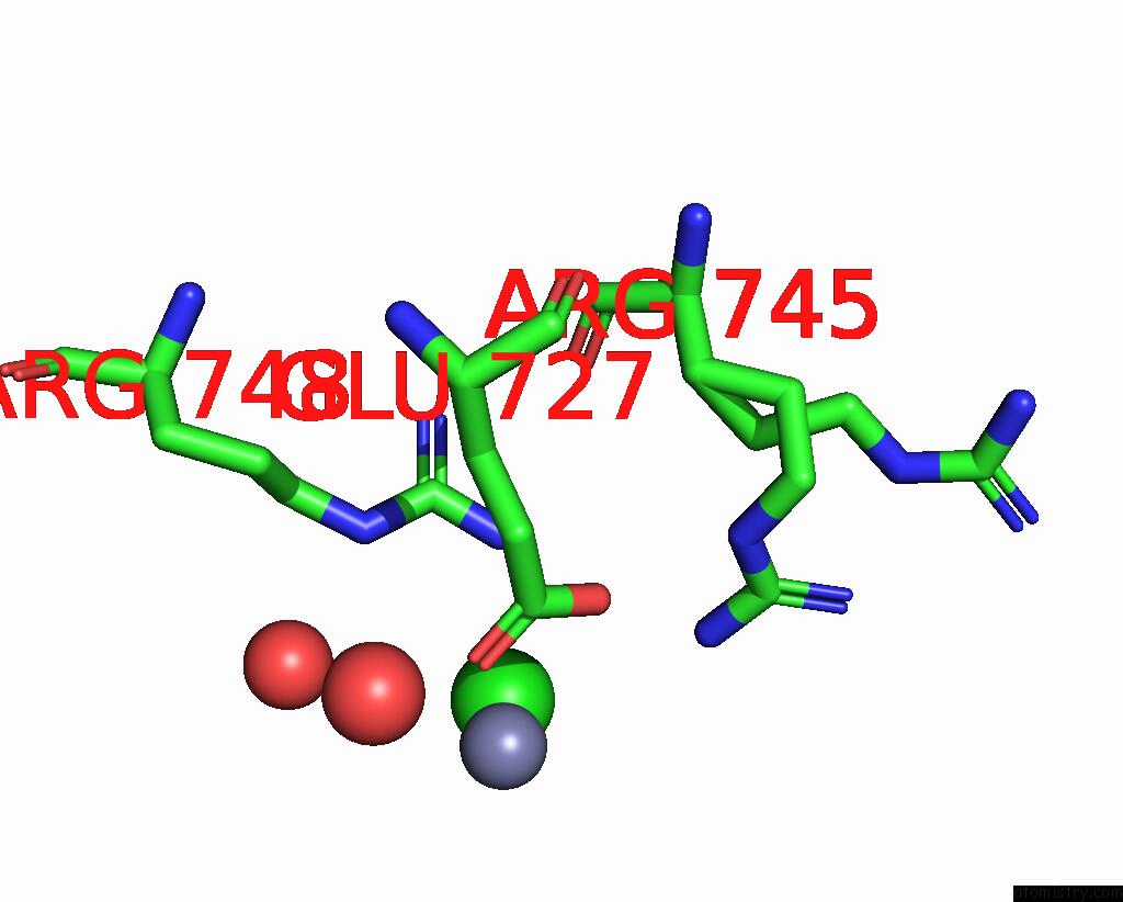
Mono view
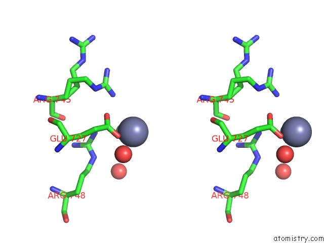
Stereo pair view
Mono view
Stereo pair view
A full contact list of Zinc with other atoms in the Zn binding
site number 2 of Crystal Structure of the Ligase Domain From M. Tuberculosis Ligd at 2.4A within 5.0Å range:
|
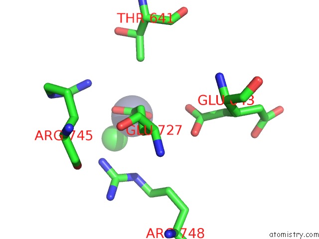
Zinc binding site 3 out of 4 in 1vs0
Go back to
Zinc binding site 3 out
of 4 in the Crystal Structure of the Ligase Domain From M. Tuberculosis Ligd at 2.4A
Mono view
Stereo pair view
Mono view
Stereo pair view
A full contact list of Zinc with other atoms in the Zn binding
site number 3 of Crystal Structure of the Ligase Domain From M. Tuberculosis Ligd at 2.4A within 5.0Å range:
|
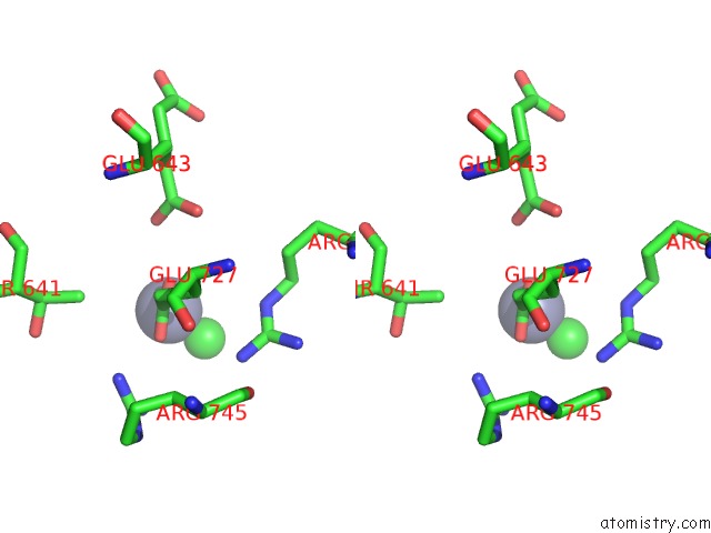
Zinc binding site 4 out of 4 in 1vs0
Go back to
Zinc binding site 4 out
of 4 in the Crystal Structure of the Ligase Domain From M. Tuberculosis Ligd at 2.4A
Mono view
Stereo pair view
Mono view
Stereo pair view
A full contact list of Zinc with other atoms in the Zn binding
site number 4 of Crystal Structure of the Ligase Domain From M. Tuberculosis Ligd at 2.4A within 5.0Å range:
|
Reference:
D.Akey,
A.Martins,
J.Aniukwu,
M.S.Glickman,
S.Shuman,
J.M.Berger.
Crystal Structure and Nonhomologous End-Joining Function of the Ligase Component of Mycobacterium Dna Ligase D. J.Biol.Chem. V. 281 13412 2006.
ISSN: ISSN 0021-9258
PubMed: 16476729
DOI: 10.1074/JBC.M513550200
Page generated: Wed Oct 16 19:52:29 2024
ISSN: ISSN 0021-9258
PubMed: 16476729
DOI: 10.1074/JBC.M513550200
Last articles
Zn in 9MJ5Zn in 9HNW
Zn in 9G0L
Zn in 9FNE
Zn in 9DZN
Zn in 9E0I
Zn in 9D32
Zn in 9DAK
Zn in 8ZXC
Zn in 8ZUF