Zinc »
PDB 1vev-1w57 »
1vje »
Zinc in PDB 1vje: Crystal Structure of A Autoinducer-2 Synthesis Protein with Bound Selenomethionine
Protein crystallography data
The structure of Crystal Structure of A Autoinducer-2 Synthesis Protein with Bound Selenomethionine, PDB code: 1vje
was solved by
Structural Genomix,
with X-Ray Crystallography technique. A brief refinement statistics is given in the table below:
| Resolution Low / High (Å) | 36.04 / 1.64 |
| Space group | P 1 21 1 |
| Cell size a, b, c (Å), α, β, γ (°) | 43.548, 81.917, 49.327, 90.00, 102.81, 90.00 |
| R / Rfree (%) | 18.8 / 21.5 |
Zinc Binding Sites:
The binding sites of Zinc atom in the Crystal Structure of A Autoinducer-2 Synthesis Protein with Bound Selenomethionine
(pdb code 1vje). This binding sites where shown within
5.0 Angstroms radius around Zinc atom.
In total 2 binding sites of Zinc where determined in the Crystal Structure of A Autoinducer-2 Synthesis Protein with Bound Selenomethionine, PDB code: 1vje:
Jump to Zinc binding site number: 1; 2;
In total 2 binding sites of Zinc where determined in the Crystal Structure of A Autoinducer-2 Synthesis Protein with Bound Selenomethionine, PDB code: 1vje:
Jump to Zinc binding site number: 1; 2;
Zinc binding site 1 out of 2 in 1vje
Go back to
Zinc binding site 1 out
of 2 in the Crystal Structure of A Autoinducer-2 Synthesis Protein with Bound Selenomethionine
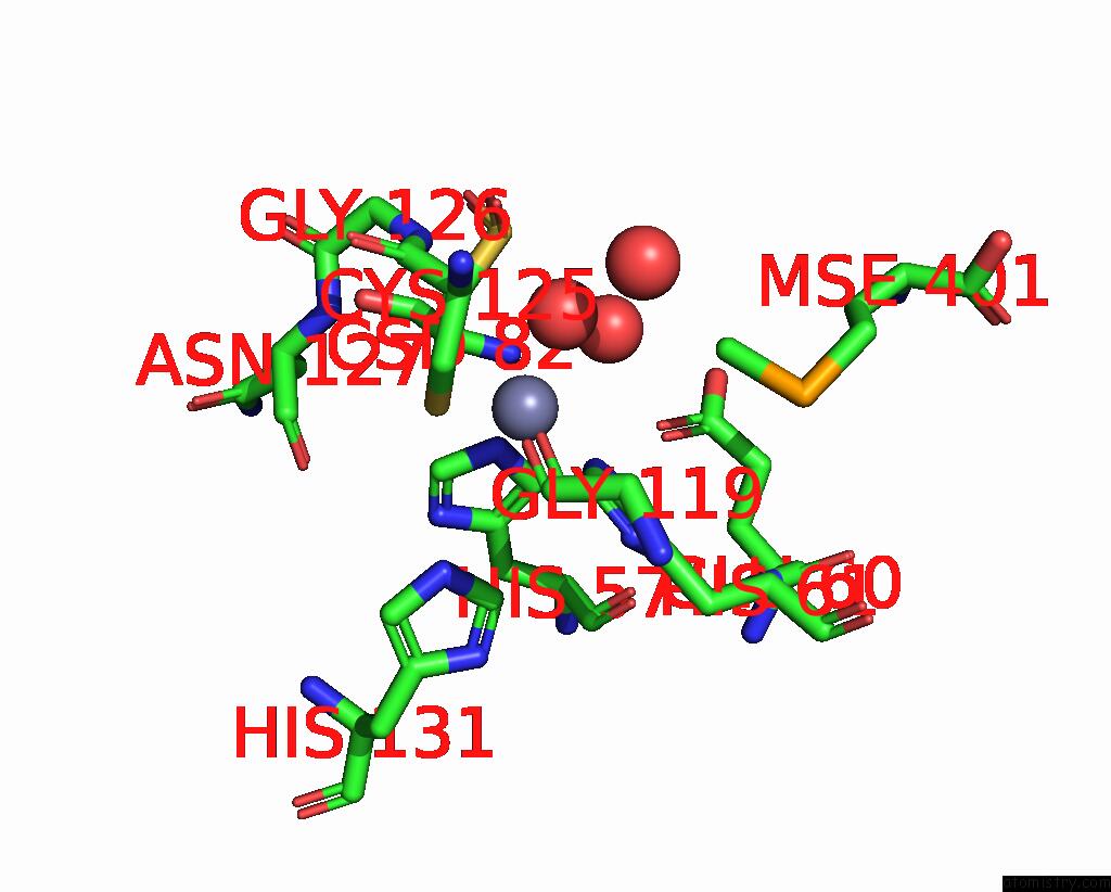
Mono view
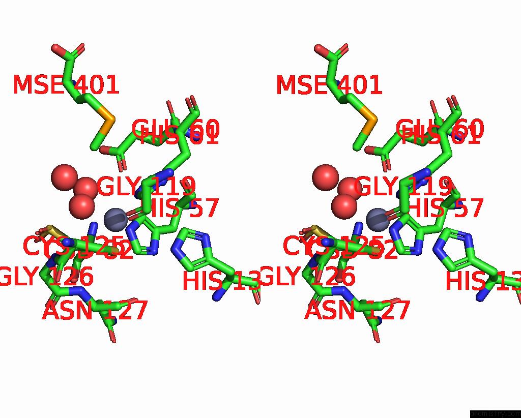
Stereo pair view
Mono view
Stereo pair view
A full contact list of Zinc with other atoms in the Zn binding
site number 1 of Crystal Structure of A Autoinducer-2 Synthesis Protein with Bound Selenomethionine within 5.0Å range:
|
Zinc binding site 2 out of 2 in 1vje
Go back to
Zinc binding site 2 out
of 2 in the Crystal Structure of A Autoinducer-2 Synthesis Protein with Bound Selenomethionine
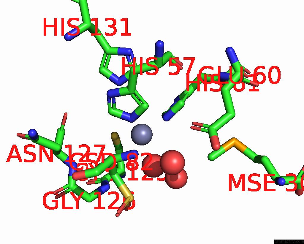
Mono view
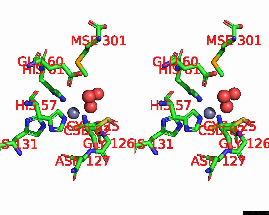
Stereo pair view
Mono view
Stereo pair view
A full contact list of Zinc with other atoms in the Zn binding
site number 2 of Crystal Structure of A Autoinducer-2 Synthesis Protein with Bound Selenomethionine within 5.0Å range:
|
Reference:
H.A.Lewis,
E.B.Furlong,
B.Laubert,
G.A.Eroshkina,
Y.Batiyenko,
J.M.Adams,
M.G.Bergseid,
C.D.Marsh,
T.S.Peat,
W.E.Sanderson,
J.M.Sauder,
S.G.Buchanan.
A Structural Genomics Approach to the Study of Quorum Sensing: Crystal Structures of Three Luxs Orthologs Structure V. 9 527 2001.
ISSN: ISSN 0969-2126
PubMed: 11435117
DOI: 10.1016/S0969-2126(01)00613-X
Page generated: Wed Oct 16 19:50:20 2024
ISSN: ISSN 0969-2126
PubMed: 11435117
DOI: 10.1016/S0969-2126(01)00613-X
Last articles
Zn in 9MJ5Zn in 9HNW
Zn in 9G0L
Zn in 9FNE
Zn in 9DZN
Zn in 9E0I
Zn in 9D32
Zn in 9DAK
Zn in 8ZXC
Zn in 8ZUF