Zinc »
PDB 1v15-1vec »
1vag »
Zinc in PDB 1vag: Neuronal Nitric Oxide Synthase Oxygenase Domain Complexed with the Inhibitor Ar-R17477
Enzymatic activity of Neuronal Nitric Oxide Synthase Oxygenase Domain Complexed with the Inhibitor Ar-R17477
All present enzymatic activity of Neuronal Nitric Oxide Synthase Oxygenase Domain Complexed with the Inhibitor Ar-R17477:
1.14.13.39;
1.14.13.39;
Protein crystallography data
The structure of Neuronal Nitric Oxide Synthase Oxygenase Domain Complexed with the Inhibitor Ar-R17477, PDB code: 1vag
was solved by
R.Fedorov,
R.Vasan,
D.K.Ghosh,
I.Schlichting,
with X-Ray Crystallography technique. A brief refinement statistics is given in the table below:
| Resolution Low / High (Å) | 8.00 / 2.00 |
| Space group | C 2 2 21 |
| Cell size a, b, c (Å), α, β, γ (°) | 45.269, 109.199, 164.885, 90.00, 90.00, 90.00 |
| R / Rfree (%) | 19.3 / 22.9 |
Other elements in 1vag:
The structure of Neuronal Nitric Oxide Synthase Oxygenase Domain Complexed with the Inhibitor Ar-R17477 also contains other interesting chemical elements:
| Iron | (Fe) | 1 atom |
| Chlorine | (Cl) | 1 atom |
Zinc Binding Sites:
The binding sites of Zinc atom in the Neuronal Nitric Oxide Synthase Oxygenase Domain Complexed with the Inhibitor Ar-R17477
(pdb code 1vag). This binding sites where shown within
5.0 Angstroms radius around Zinc atom.
In total only one binding site of Zinc was determined in the Neuronal Nitric Oxide Synthase Oxygenase Domain Complexed with the Inhibitor Ar-R17477, PDB code: 1vag:
In total only one binding site of Zinc was determined in the Neuronal Nitric Oxide Synthase Oxygenase Domain Complexed with the Inhibitor Ar-R17477, PDB code: 1vag:
Zinc binding site 1 out of 1 in 1vag
Go back to
Zinc binding site 1 out
of 1 in the Neuronal Nitric Oxide Synthase Oxygenase Domain Complexed with the Inhibitor Ar-R17477
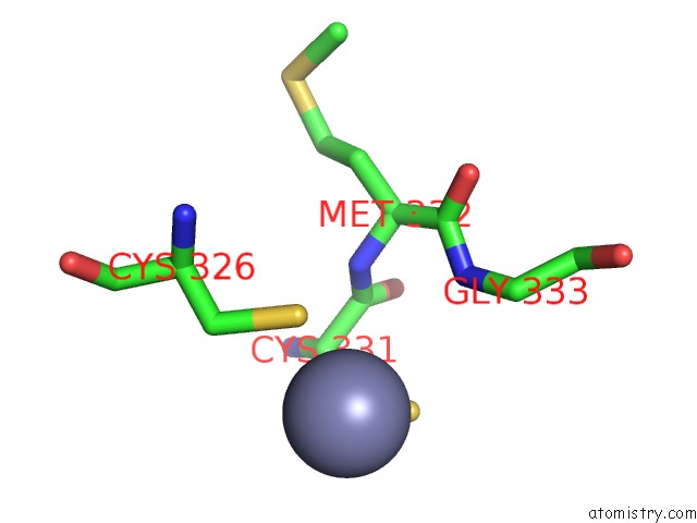
Mono view
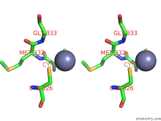
Stereo pair view
Mono view
Stereo pair view
A full contact list of Zinc with other atoms in the Zn binding
site number 1 of Neuronal Nitric Oxide Synthase Oxygenase Domain Complexed with the Inhibitor Ar-R17477 within 5.0Å range:
|
Reference:
R.Fedorov,
R.Vasan,
D.K.Ghosh,
I.Schlichting.
Structures of Nitric Oxide Synthase Isoforms Complexed with the Inhibitor Ar-R17477 Suggest A Rational Basis For Specificity and Inhibitor Design Proc.Natl.Acad.Sci.Usa V. 101 5892 2004.
ISSN: ISSN 0027-8424
PubMed: 15071192
DOI: 10.1073/PNAS.0306588101
Page generated: Wed Oct 16 19:47:23 2024
ISSN: ISSN 0027-8424
PubMed: 15071192
DOI: 10.1073/PNAS.0306588101
Last articles
Zn in 9MJ5Zn in 9HNW
Zn in 9G0L
Zn in 9FNE
Zn in 9DZN
Zn in 9E0I
Zn in 9D32
Zn in 9DAK
Zn in 8ZXC
Zn in 8ZUF