Zinc »
PDB 1ul4-1v13 »
1uol »
Zinc in PDB 1uol: Crystal Structure of the Human P53 Core Domain Mutant M133L/V203A/N239Y/N268D at 1.9 A Resolution.
Protein crystallography data
The structure of Crystal Structure of the Human P53 Core Domain Mutant M133L/V203A/N239Y/N268D at 1.9 A Resolution., PDB code: 1uol
was solved by
A.C.Joerger,
M.D.Allen,
A.R.Fersht,
with X-Ray Crystallography technique. A brief refinement statistics is given in the table below:
| Resolution Low / High (Å) | 35.6 / 1.9 |
| Space group | P 21 21 21 |
| Cell size a, b, c (Å), α, β, γ (°) | 65.000, 71.000, 104.800, 90.00, 90.00, 90.00 |
| R / Rfree (%) | 19.2 / 23 |
Zinc Binding Sites:
The binding sites of Zinc atom in the Crystal Structure of the Human P53 Core Domain Mutant M133L/V203A/N239Y/N268D at 1.9 A Resolution.
(pdb code 1uol). This binding sites where shown within
5.0 Angstroms radius around Zinc atom.
In total 2 binding sites of Zinc where determined in the Crystal Structure of the Human P53 Core Domain Mutant M133L/V203A/N239Y/N268D at 1.9 A Resolution., PDB code: 1uol:
Jump to Zinc binding site number: 1; 2;
In total 2 binding sites of Zinc where determined in the Crystal Structure of the Human P53 Core Domain Mutant M133L/V203A/N239Y/N268D at 1.9 A Resolution., PDB code: 1uol:
Jump to Zinc binding site number: 1; 2;
Zinc binding site 1 out of 2 in 1uol
Go back to
Zinc binding site 1 out
of 2 in the Crystal Structure of the Human P53 Core Domain Mutant M133L/V203A/N239Y/N268D at 1.9 A Resolution.
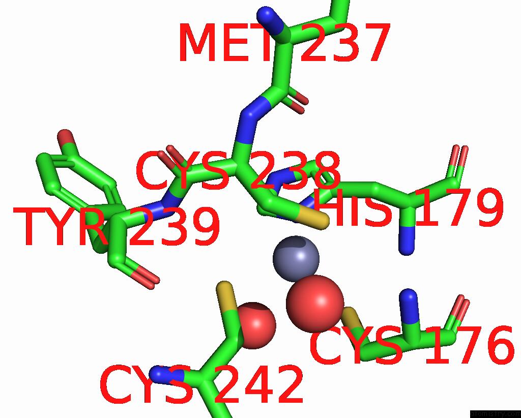
Mono view
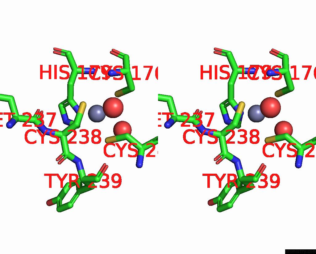
Stereo pair view
Mono view
Stereo pair view
A full contact list of Zinc with other atoms in the Zn binding
site number 1 of Crystal Structure of the Human P53 Core Domain Mutant M133L/V203A/N239Y/N268D at 1.9 A Resolution. within 5.0Å range:
|
Zinc binding site 2 out of 2 in 1uol
Go back to
Zinc binding site 2 out
of 2 in the Crystal Structure of the Human P53 Core Domain Mutant M133L/V203A/N239Y/N268D at 1.9 A Resolution.
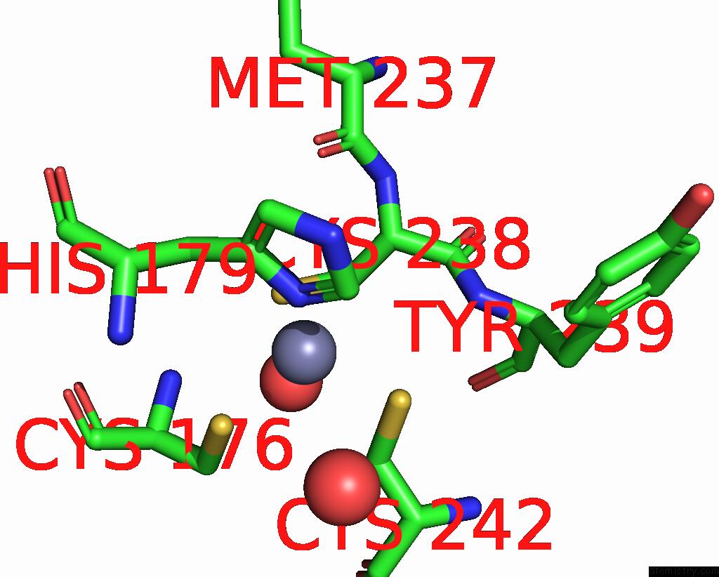
Mono view
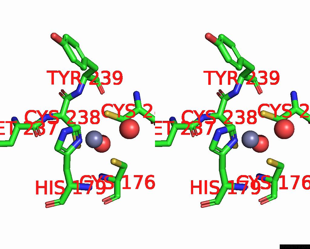
Stereo pair view
Mono view
Stereo pair view
A full contact list of Zinc with other atoms in the Zn binding
site number 2 of Crystal Structure of the Human P53 Core Domain Mutant M133L/V203A/N239Y/N268D at 1.9 A Resolution. within 5.0Å range:
|
Reference:
A.C.Joerger,
M.D.Allen,
A.R.Fersht.
Crystal Structure of A Superstable Mutant of Human P53 Core Domain. Insights Into the Mechanism of Rescuing Oncogenic Mutations J.Biol.Chem. V. 279 1291 2004.
ISSN: ISSN 0021-9258
PubMed: 14534297
DOI: 10.1074/JBC.M309732200
Page generated: Wed Oct 16 19:34:26 2024
ISSN: ISSN 0021-9258
PubMed: 14534297
DOI: 10.1074/JBC.M309732200
Last articles
Zn in 9MJ5Zn in 9HNW
Zn in 9G0L
Zn in 9FNE
Zn in 9DZN
Zn in 9E0I
Zn in 9D32
Zn in 9DAK
Zn in 8ZXC
Zn in 8ZUF