Zinc »
PDB 1u2n-1uip »
1u7j »
Zinc in PDB 1u7j: Solution Structure of A Diiron Protein Model
Zinc Binding Sites:
The binding sites of Zinc atom in the Solution Structure of A Diiron Protein Model
(pdb code 1u7j). This binding sites where shown within
5.0 Angstroms radius around Zinc atom.
In total 2 binding sites of Zinc where determined in the Solution Structure of A Diiron Protein Model, PDB code: 1u7j:
Jump to Zinc binding site number: 1; 2;
In total 2 binding sites of Zinc where determined in the Solution Structure of A Diiron Protein Model, PDB code: 1u7j:
Jump to Zinc binding site number: 1; 2;
Zinc binding site 1 out of 2 in 1u7j
Go back to
Zinc binding site 1 out
of 2 in the Solution Structure of A Diiron Protein Model
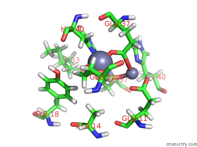
Mono view
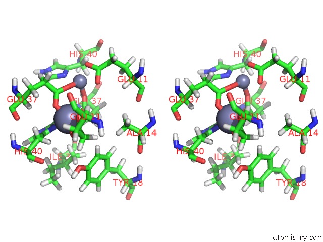
Stereo pair view
Mono view
Stereo pair view
A full contact list of Zinc with other atoms in the Zn binding
site number 1 of Solution Structure of A Diiron Protein Model within 5.0Å range:
|
Zinc binding site 2 out of 2 in 1u7j
Go back to
Zinc binding site 2 out
of 2 in the Solution Structure of A Diiron Protein Model
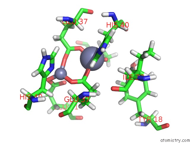
Mono view
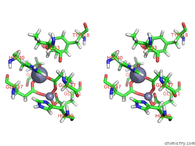
Stereo pair view
Mono view
Stereo pair view
A full contact list of Zinc with other atoms in the Zn binding
site number 2 of Solution Structure of A Diiron Protein Model within 5.0Å range:
|
Reference:
S.J.Lahr,
D.E.Engel,
S.E.Stayrook,
O.Maglio,
B.North,
S.Geremia,
A.Lombardi,
W.F.Degrado.
Analysis and Design of Turns in Alpha-Helical Hairpins J.Mol.Biol. V. 346 1441 2005.
ISSN: ISSN 0022-2836
PubMed: 15713492
DOI: 10.1016/J.JMB.2004.12.016
Page generated: Wed Oct 16 19:28:30 2024
ISSN: ISSN 0022-2836
PubMed: 15713492
DOI: 10.1016/J.JMB.2004.12.016
Last articles
Zn in 9MJ5Zn in 9HNW
Zn in 9G0L
Zn in 9FNE
Zn in 9DZN
Zn in 9E0I
Zn in 9D32
Zn in 9DAK
Zn in 8ZXC
Zn in 8ZUF