Zinc »
PDB 1to5-1u22 »
1tu0 »
Zinc in PDB 1tu0: Aspartate Transcarbamoylase Catalytic Chain Mutant E50A Complex with Phosphonoacetamide
Enzymatic activity of Aspartate Transcarbamoylase Catalytic Chain Mutant E50A Complex with Phosphonoacetamide
All present enzymatic activity of Aspartate Transcarbamoylase Catalytic Chain Mutant E50A Complex with Phosphonoacetamide:
2.1.3.2;
2.1.3.2;
Protein crystallography data
The structure of Aspartate Transcarbamoylase Catalytic Chain Mutant E50A Complex with Phosphonoacetamide, PDB code: 1tu0
was solved by
K.Stieglitz,
B.Stec,
D.P.Baker,
E.R.Kantrowitz,
with X-Ray Crystallography technique. A brief refinement statistics is given in the table below:
| Resolution Low / High (Å) | 30.00 / 2.55 |
| Space group | P 3 2 1 |
| Cell size a, b, c (Å), α, β, γ (°) | 122.200, 122.200, 142.700, 90.00, 90.00, 120.00 |
| R / Rfree (%) | 19 / 27.9 |
Zinc Binding Sites:
The binding sites of Zinc atom in the Aspartate Transcarbamoylase Catalytic Chain Mutant E50A Complex with Phosphonoacetamide
(pdb code 1tu0). This binding sites where shown within
5.0 Angstroms radius around Zinc atom.
In total 2 binding sites of Zinc where determined in the Aspartate Transcarbamoylase Catalytic Chain Mutant E50A Complex with Phosphonoacetamide, PDB code: 1tu0:
Jump to Zinc binding site number: 1; 2;
In total 2 binding sites of Zinc where determined in the Aspartate Transcarbamoylase Catalytic Chain Mutant E50A Complex with Phosphonoacetamide, PDB code: 1tu0:
Jump to Zinc binding site number: 1; 2;
Zinc binding site 1 out of 2 in 1tu0
Go back to
Zinc binding site 1 out
of 2 in the Aspartate Transcarbamoylase Catalytic Chain Mutant E50A Complex with Phosphonoacetamide
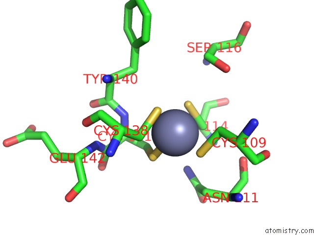
Mono view
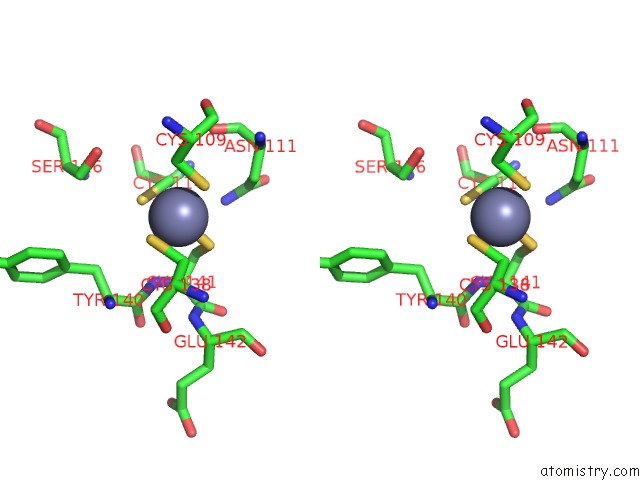
Stereo pair view
Mono view
Stereo pair view
A full contact list of Zinc with other atoms in the Zn binding
site number 1 of Aspartate Transcarbamoylase Catalytic Chain Mutant E50A Complex with Phosphonoacetamide within 5.0Å range:
|
Zinc binding site 2 out of 2 in 1tu0
Go back to
Zinc binding site 2 out
of 2 in the Aspartate Transcarbamoylase Catalytic Chain Mutant E50A Complex with Phosphonoacetamide
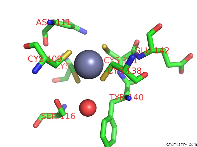
Mono view
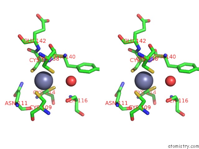
Stereo pair view
Mono view
Stereo pair view
A full contact list of Zinc with other atoms in the Zn binding
site number 2 of Aspartate Transcarbamoylase Catalytic Chain Mutant E50A Complex with Phosphonoacetamide within 5.0Å range:
|
Reference:
K.Stieglitz,
B.Stec,
D.P.Baker,
E.R.Kantrowitz.
Monitoring the Transition From the T to the R State in E.Coli Aspartate Transcarbamoylase By X-Ray Crystallography: Crystal Structures of the E50A Mutant Enzyme in Four Distinct Allosteric States. J.Mol.Biol. V. 341 853 2004.
ISSN: ISSN 0022-2836
PubMed: 15288791
DOI: 10.1016/J.JMB.2004.06.002
Page generated: Wed Oct 16 19:17:53 2024
ISSN: ISSN 0022-2836
PubMed: 15288791
DOI: 10.1016/J.JMB.2004.06.002
Last articles
Zn in 9MJ5Zn in 9HNW
Zn in 9G0L
Zn in 9FNE
Zn in 9DZN
Zn in 9E0I
Zn in 9D32
Zn in 9DAK
Zn in 8ZXC
Zn in 8ZUF