Zinc »
PDB 1to5-1u22 »
1to5 »
Zinc in PDB 1to5: Structure of the Cytosolic Cu,Zn Sod From S. Mansoni
Enzymatic activity of Structure of the Cytosolic Cu,Zn Sod From S. Mansoni
All present enzymatic activity of Structure of the Cytosolic Cu,Zn Sod From S. Mansoni:
1.15.1.1;
1.15.1.1;
Protein crystallography data
The structure of Structure of the Cytosolic Cu,Zn Sod From S. Mansoni, PDB code: 1to5
was solved by
R.M.F.Cardoso,
C.H.T.P.Silva,
A.P.Ulian De Araujo,
T.Tanaka,
M.Tanaka,
R.C.Garratt,
with X-Ray Crystallography technique. A brief refinement statistics is given in the table below:
| Resolution Low / High (Å) | 58.80 / 2.20 |
| Space group | P 21 21 21 |
| Cell size a, b, c (Å), α, β, γ (°) | 74.640, 78.240, 95.180, 90.00, 90.00, 90.00 |
| R / Rfree (%) | 17.6 / 24.1 |
Other elements in 1to5:
The structure of Structure of the Cytosolic Cu,Zn Sod From S. Mansoni also contains other interesting chemical elements:
| Copper | (Cu) | 4 atoms |
Zinc Binding Sites:
The binding sites of Zinc atom in the Structure of the Cytosolic Cu,Zn Sod From S. Mansoni
(pdb code 1to5). This binding sites where shown within
5.0 Angstroms radius around Zinc atom.
In total 4 binding sites of Zinc where determined in the Structure of the Cytosolic Cu,Zn Sod From S. Mansoni, PDB code: 1to5:
Jump to Zinc binding site number: 1; 2; 3; 4;
In total 4 binding sites of Zinc where determined in the Structure of the Cytosolic Cu,Zn Sod From S. Mansoni, PDB code: 1to5:
Jump to Zinc binding site number: 1; 2; 3; 4;
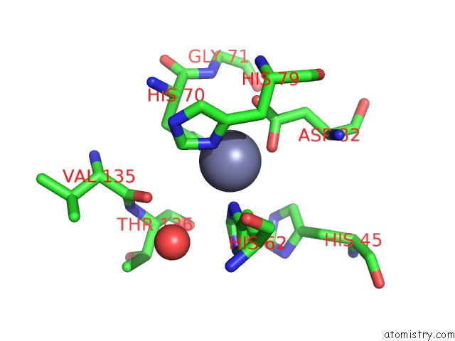
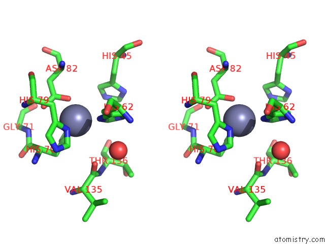
Zinc binding site 1 out of 4 in 1to5
Go back to
Zinc binding site 1 out
of 4 in the Structure of the Cytosolic Cu,Zn Sod From S. Mansoni
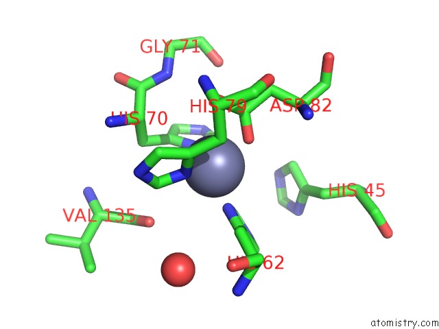
Mono view
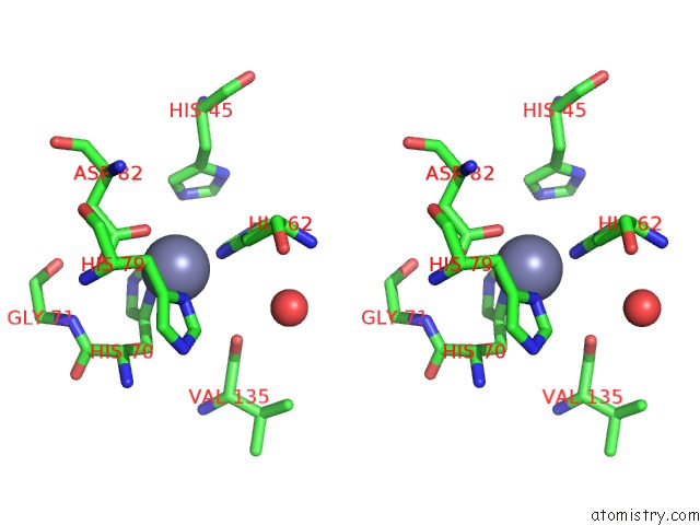
Stereo pair view
Mono view
Stereo pair view
A full contact list of Zinc with other atoms in the Zn binding
site number 1 of Structure of the Cytosolic Cu,Zn Sod From S. Mansoni within 5.0Å range:
|
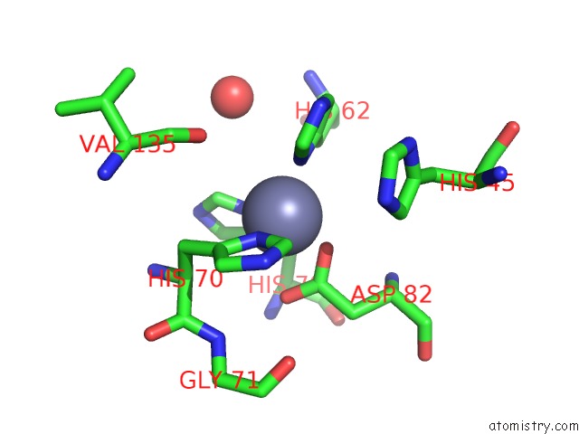
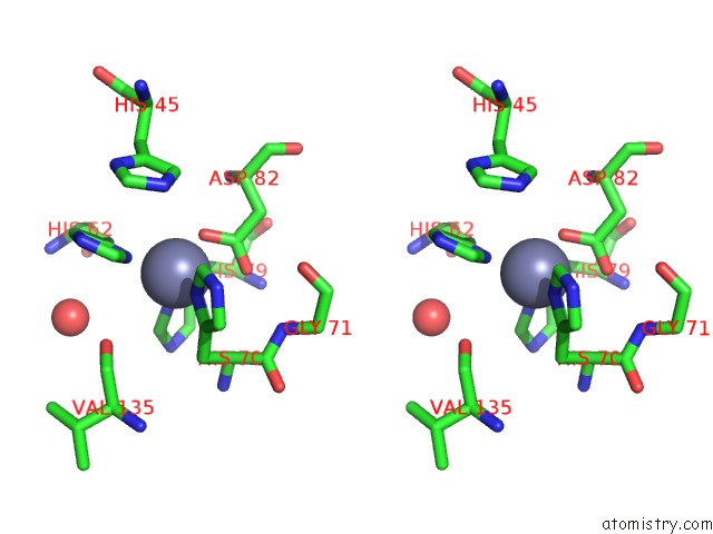
Zinc binding site 2 out of 4 in 1to5
Go back to
Zinc binding site 2 out
of 4 in the Structure of the Cytosolic Cu,Zn Sod From S. Mansoni
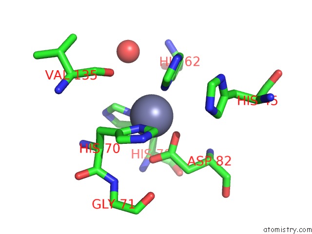
Mono view
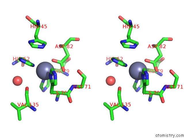
Stereo pair view
Mono view
Stereo pair view
A full contact list of Zinc with other atoms in the Zn binding
site number 2 of Structure of the Cytosolic Cu,Zn Sod From S. Mansoni within 5.0Å range:
|
Zinc binding site 3 out of 4 in 1to5
Go back to
Zinc binding site 3 out
of 4 in the Structure of the Cytosolic Cu,Zn Sod From S. Mansoni
Mono view
Stereo pair view
Mono view
Stereo pair view
A full contact list of Zinc with other atoms in the Zn binding
site number 3 of Structure of the Cytosolic Cu,Zn Sod From S. Mansoni within 5.0Å range:
|
Zinc binding site 4 out of 4 in 1to5
Go back to
Zinc binding site 4 out
of 4 in the Structure of the Cytosolic Cu,Zn Sod From S. Mansoni
Mono view
Stereo pair view
Mono view
Stereo pair view
A full contact list of Zinc with other atoms in the Zn binding
site number 4 of Structure of the Cytosolic Cu,Zn Sod From S. Mansoni within 5.0Å range:
|
Reference:
R.M.Cardoso,
C.H.Silva,
A.P.Ulian De Araujo,
T.Tanaka,
M.Tanaka,
R.C.Garratt.
Structure of the Cytosolic Cu,Zn Superoxide Dismutase From Schistosoma Mansoni. Acta Crystallogr.,Sect.D V. 60 1569 2004.
ISSN: ISSN 0907-4449
PubMed: 15333927
DOI: 10.1107/S0907444904016798
Page generated: Wed Oct 16 19:14:35 2024
ISSN: ISSN 0907-4449
PubMed: 15333927
DOI: 10.1107/S0907444904016798
Last articles
As in 2VDLAs in 2VEQ
As in 2VDK
As in 2V5C
As in 2V96
As in 2RA8
As in 2PQ3
As in 2OUI
As in 2RL7
As in 2O4Q