Zinc »
PDB 1tbt-1to4 »
1tdz »
Zinc in PDB 1tdz: Crystal Structure Complex Between the Lactococcus Lactis Fpg (Mutm) and A Fapy-Dg Containing Dna
Enzymatic activity of Crystal Structure Complex Between the Lactococcus Lactis Fpg (Mutm) and A Fapy-Dg Containing Dna
All present enzymatic activity of Crystal Structure Complex Between the Lactococcus Lactis Fpg (Mutm) and A Fapy-Dg Containing Dna:
3.2.2.23;
3.2.2.23;
Protein crystallography data
The structure of Crystal Structure Complex Between the Lactococcus Lactis Fpg (Mutm) and A Fapy-Dg Containing Dna, PDB code: 1tdz
was solved by
F.Coste,
M.Ober,
T.Carell,
S.Boiteux,
C.Zelwer,
B.Castaing,
with X-Ray Crystallography technique. A brief refinement statistics is given in the table below:
| Resolution Low / High (Å) | 20.00 / 1.80 |
| Space group | P 41 21 2 |
| Cell size a, b, c (Å), α, β, γ (°) | 91.408, 91.408, 141.575, 90.00, 90.00, 90.00 |
| R / Rfree (%) | 17.9 / 20.7 |
Zinc Binding Sites:
The binding sites of Zinc atom in the Crystal Structure Complex Between the Lactococcus Lactis Fpg (Mutm) and A Fapy-Dg Containing Dna
(pdb code 1tdz). This binding sites where shown within
5.0 Angstroms radius around Zinc atom.
In total only one binding site of Zinc was determined in the Crystal Structure Complex Between the Lactococcus Lactis Fpg (Mutm) and A Fapy-Dg Containing Dna, PDB code: 1tdz:
In total only one binding site of Zinc was determined in the Crystal Structure Complex Between the Lactococcus Lactis Fpg (Mutm) and A Fapy-Dg Containing Dna, PDB code: 1tdz:
Zinc binding site 1 out of 1 in 1tdz
Go back to
Zinc binding site 1 out
of 1 in the Crystal Structure Complex Between the Lactococcus Lactis Fpg (Mutm) and A Fapy-Dg Containing Dna
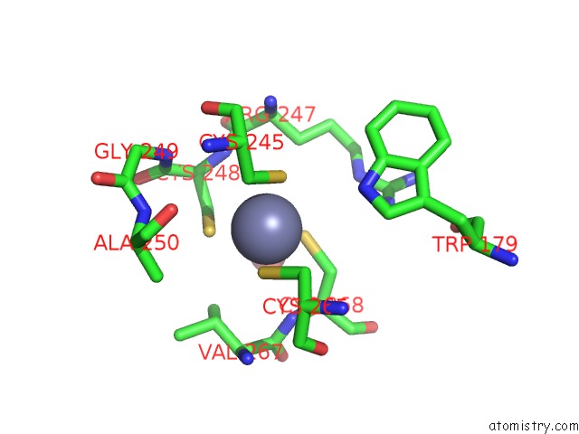
Mono view
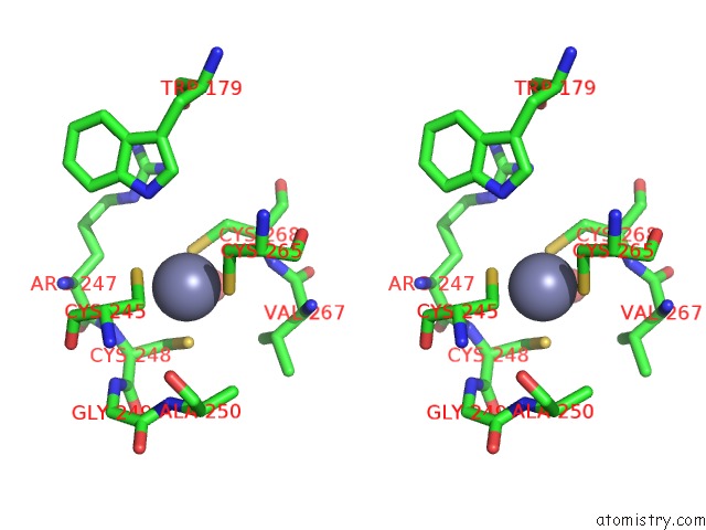
Stereo pair view
Mono view
Stereo pair view
A full contact list of Zinc with other atoms in the Zn binding
site number 1 of Crystal Structure Complex Between the Lactococcus Lactis Fpg (Mutm) and A Fapy-Dg Containing Dna within 5.0Å range:
|
Reference:
F.Coste,
M.Ober,
T.Carell,
S.Boiteux,
C.Zelwer,
B.Castaing.
Structural Basis For the Recognition of the Fapydg Lesion (2,6-Diamino-4-Hydroxy-5-Formamidopyrimidine) By Formamidopyrimidine-Dna Glycosylase J.Biol.Chem. V. 279 44074 2004.
ISSN: ISSN 0021-9258
PubMed: 15249553
DOI: 10.1074/JBC.M405928200
Page generated: Tue Aug 19 23:19:07 2025
ISSN: ISSN 0021-9258
PubMed: 15249553
DOI: 10.1074/JBC.M405928200
Last articles
Zn in 2APSZn in 2AQC
Zn in 2AQ2
Zn in 2APO
Zn in 2AN6
Zn in 2AP1
Zn in 2ANH
Zn in 2ANP
Zn in 2AMT
Zn in 2AL4