Zinc »
PDB 1sxc-1tbo »
1sxc »
Zinc in PDB 1sxc: Crystal Structure of Reduced Bovine Erythrocyte Superoxide Dismutase at 1.9 Angstroms Resolution
Enzymatic activity of Crystal Structure of Reduced Bovine Erythrocyte Superoxide Dismutase at 1.9 Angstroms Resolution
All present enzymatic activity of Crystal Structure of Reduced Bovine Erythrocyte Superoxide Dismutase at 1.9 Angstroms Resolution:
1.15.1.1;
1.15.1.1;
Protein crystallography data
The structure of Crystal Structure of Reduced Bovine Erythrocyte Superoxide Dismutase at 1.9 Angstroms Resolution, PDB code: 1sxc
was solved by
W.R.Rypniewski,
S.Mangani,
B.Bruni,
P.Orioli,
M.Casati,
K.S.Wilson,
with X-Ray Crystallography technique. A brief refinement statistics is given in the table below:
| Resolution Low / High (Å) | 10.00 / 1.90 |
| Space group | P 21 21 21 |
| Cell size a, b, c (Å), α, β, γ (°) | 47.800, 51.060, 147.990, 90.00, 90.00, 90.00 |
| R / Rfree (%) | n/a / n/a |
Other elements in 1sxc:
The structure of Crystal Structure of Reduced Bovine Erythrocyte Superoxide Dismutase at 1.9 Angstroms Resolution also contains other interesting chemical elements:
| Copper | (Cu) | 2 atoms |
Zinc Binding Sites:
The binding sites of Zinc atom in the Crystal Structure of Reduced Bovine Erythrocyte Superoxide Dismutase at 1.9 Angstroms Resolution
(pdb code 1sxc). This binding sites where shown within
5.0 Angstroms radius around Zinc atom.
In total 2 binding sites of Zinc where determined in the Crystal Structure of Reduced Bovine Erythrocyte Superoxide Dismutase at 1.9 Angstroms Resolution, PDB code: 1sxc:
Jump to Zinc binding site number: 1; 2;
In total 2 binding sites of Zinc where determined in the Crystal Structure of Reduced Bovine Erythrocyte Superoxide Dismutase at 1.9 Angstroms Resolution, PDB code: 1sxc:
Jump to Zinc binding site number: 1; 2;
Zinc binding site 1 out of 2 in 1sxc
Go back to
Zinc binding site 1 out
of 2 in the Crystal Structure of Reduced Bovine Erythrocyte Superoxide Dismutase at 1.9 Angstroms Resolution
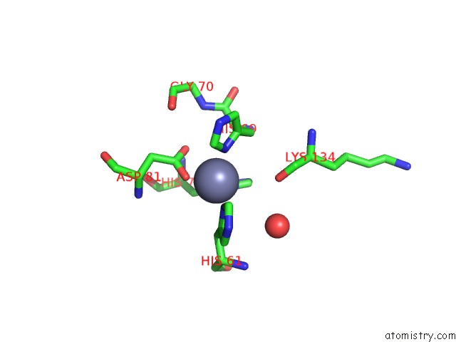
Mono view
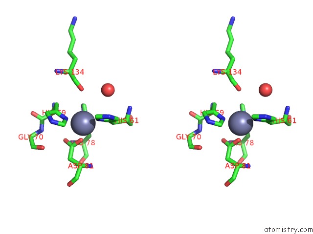
Stereo pair view
Mono view
Stereo pair view
A full contact list of Zinc with other atoms in the Zn binding
site number 1 of Crystal Structure of Reduced Bovine Erythrocyte Superoxide Dismutase at 1.9 Angstroms Resolution within 5.0Å range:
|
Zinc binding site 2 out of 2 in 1sxc
Go back to
Zinc binding site 2 out
of 2 in the Crystal Structure of Reduced Bovine Erythrocyte Superoxide Dismutase at 1.9 Angstroms Resolution
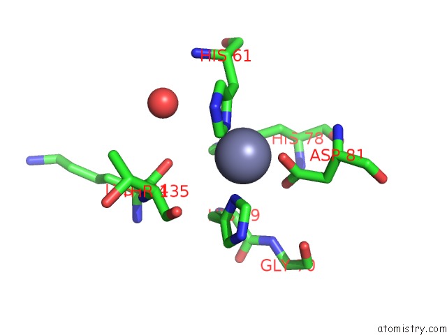
Mono view

Stereo pair view
Mono view
Stereo pair view
A full contact list of Zinc with other atoms in the Zn binding
site number 2 of Crystal Structure of Reduced Bovine Erythrocyte Superoxide Dismutase at 1.9 Angstroms Resolution within 5.0Å range:
|
Reference:
W.R.Rypniewski,
S.Mangani,
B.Bruni,
P.L.Orioli,
M.Casati,
K.S.Wilson.
Crystal Structure of Reduced Bovine Erythrocyte Superoxide Dismutase at 1.9 A Resolution. J.Mol.Biol. V. 251 282 1995.
ISSN: ISSN 0022-2836
PubMed: 7643403
DOI: 10.1006/JMBI.1995.0434
Page generated: Tue Aug 19 23:13:44 2025
ISSN: ISSN 0022-2836
PubMed: 7643403
DOI: 10.1006/JMBI.1995.0434
Last articles
Zn in 2ANUZn in 2AQR
Zn in 2AQQ
Zn in 2AQO
Zn in 2AQP
Zn in 2AQN
Zn in 2APS
Zn in 2AQC
Zn in 2AQ2
Zn in 2APO