Zinc »
PDB 1sdz-1sxb »
1sg0 »
Zinc in PDB 1sg0: Crystal Structure Analysis of QR2 in Complex with Resveratrol
Enzymatic activity of Crystal Structure Analysis of QR2 in Complex with Resveratrol
All present enzymatic activity of Crystal Structure Analysis of QR2 in Complex with Resveratrol:
1.6.99.2;
1.6.99.2;
Protein crystallography data
The structure of Crystal Structure Analysis of QR2 in Complex with Resveratrol, PDB code: 1sg0
was solved by
L.Buryanovskyy,
Y.Fu,
M.Boyd,
Y.Ma,
T.C.Tsieh,
J.M.Wu,
Z.Zhang,
with X-Ray Crystallography technique. A brief refinement statistics is given in the table below:
| Resolution Low / High (Å) | 28.43 / 1.50 |
| Space group | P 21 21 21 |
| Cell size a, b, c (Å), α, β, γ (°) | 83.330, 106.372, 56.982, 90.00, 90.00, 90.00 |
| R / Rfree (%) | 21.4 / 23.4 |
Zinc Binding Sites:
The binding sites of Zinc atom in the Crystal Structure Analysis of QR2 in Complex with Resveratrol
(pdb code 1sg0). This binding sites where shown within
5.0 Angstroms radius around Zinc atom.
In total 2 binding sites of Zinc where determined in the Crystal Structure Analysis of QR2 in Complex with Resveratrol, PDB code: 1sg0:
Jump to Zinc binding site number: 1; 2;
In total 2 binding sites of Zinc where determined in the Crystal Structure Analysis of QR2 in Complex with Resveratrol, PDB code: 1sg0:
Jump to Zinc binding site number: 1; 2;
Zinc binding site 1 out of 2 in 1sg0
Go back to
Zinc binding site 1 out
of 2 in the Crystal Structure Analysis of QR2 in Complex with Resveratrol
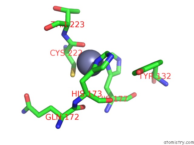
Mono view
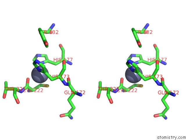
Stereo pair view
Mono view
Stereo pair view
A full contact list of Zinc with other atoms in the Zn binding
site number 1 of Crystal Structure Analysis of QR2 in Complex with Resveratrol within 5.0Å range:
|
Zinc binding site 2 out of 2 in 1sg0
Go back to
Zinc binding site 2 out
of 2 in the Crystal Structure Analysis of QR2 in Complex with Resveratrol
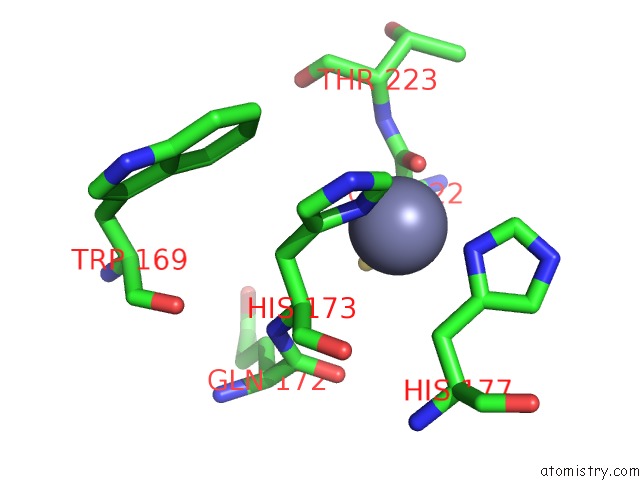
Mono view
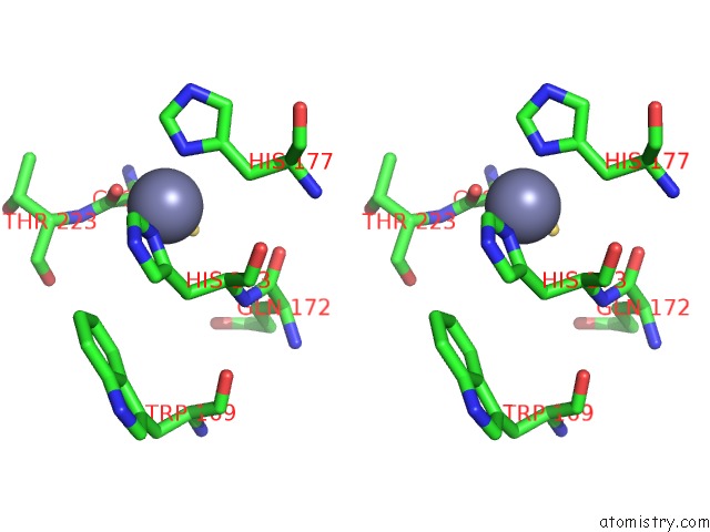
Stereo pair view
Mono view
Stereo pair view
A full contact list of Zinc with other atoms in the Zn binding
site number 2 of Crystal Structure Analysis of QR2 in Complex with Resveratrol within 5.0Å range:
|
Reference:
L.Buryanovskyy,
Y.Fu,
M.Boyd,
Y.Ma,
T.C.Hsieh,
J.M.Wu,
Z.Zhang.
Crystal Structure of Quinone Reductase 2 in Complex with Resveratrol Biochemistry V. 43 11417 2004.
ISSN: ISSN 0006-2960
PubMed: 15350128
DOI: 10.1021/BI049162O
Page generated: Tue Aug 19 23:07:23 2025
ISSN: ISSN 0006-2960
PubMed: 15350128
DOI: 10.1021/BI049162O
Last articles
Zn in 2APSZn in 2AQC
Zn in 2AQ2
Zn in 2APO
Zn in 2AN6
Zn in 2AP1
Zn in 2ANH
Zn in 2ANP
Zn in 2AMT
Zn in 2AL4