Zinc »
PDB 1rag-1rqg »
1rpj »
Zinc in PDB 1rpj: Crystal Structure of D-Allose Binding Protein From Escherichia Coli
Protein crystallography data
The structure of Crystal Structure of D-Allose Binding Protein From Escherichia Coli, PDB code: 1rpj
was solved by
B.Chaudhuri,
T.A.Jones,
S.L.Mowbray,
with X-Ray Crystallography technique. A brief refinement statistics is given in the table below:
| Resolution Low / High (Å) | 29.00 / 1.80 |
| Space group | P 1 21 1 |
| Cell size a, b, c (Å), α, β, γ (°) | 33.380, 67.820, 53.590, 90.00, 96.71, 90.00 |
| R / Rfree (%) | 19.4 / 24.5 |
Zinc Binding Sites:
The binding sites of Zinc atom in the Crystal Structure of D-Allose Binding Protein From Escherichia Coli
(pdb code 1rpj). This binding sites where shown within
5.0 Angstroms radius around Zinc atom.
In total only one binding site of Zinc was determined in the Crystal Structure of D-Allose Binding Protein From Escherichia Coli, PDB code: 1rpj:
In total only one binding site of Zinc was determined in the Crystal Structure of D-Allose Binding Protein From Escherichia Coli, PDB code: 1rpj:
Zinc binding site 1 out of 1 in 1rpj
Go back to
Zinc binding site 1 out
of 1 in the Crystal Structure of D-Allose Binding Protein From Escherichia Coli
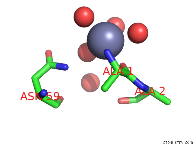
Mono view
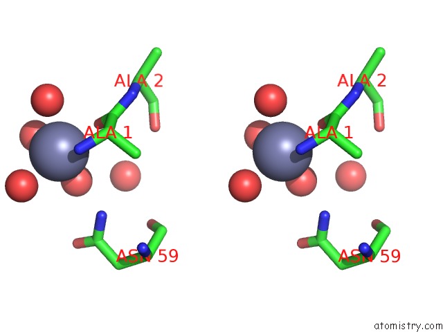
Stereo pair view
Mono view
Stereo pair view
A full contact list of Zinc with other atoms in the Zn binding
site number 1 of Crystal Structure of D-Allose Binding Protein From Escherichia Coli within 5.0Å range:
|
Reference:
B.N.Chaudhuri,
J.Ko,
C.Park,
T.A.Jones,
S.L.Mowbray.
Structure of D-Allose Binding Protein From Escherichia Coli Bound to D-Allose at 1.8 A Resolution. J.Mol.Biol. V. 286 1519 1999.
ISSN: ISSN 0022-2836
PubMed: 10064713
DOI: 10.1006/JMBI.1999.2571
Page generated: Wed Oct 16 18:39:11 2024
ISSN: ISSN 0022-2836
PubMed: 10064713
DOI: 10.1006/JMBI.1999.2571
Last articles
Zn in 9MJ5Zn in 9HNW
Zn in 9G0L
Zn in 9FNE
Zn in 9DZN
Zn in 9E0I
Zn in 9D32
Zn in 9DAK
Zn in 8ZXC
Zn in 8ZUF