Zinc »
PDB 1qmu-1r23 »
1qx2 »
Zinc in PDB 1qx2: X-Ray Structure of Calcium-Loaded Calbindomodulin (A Calbindin D9K Re- Engineered to Undergo A Conformational Opening) at 1.44 A Resolution
Protein crystallography data
The structure of X-Ray Structure of Calcium-Loaded Calbindomodulin (A Calbindin D9K Re- Engineered to Undergo A Conformational Opening) at 1.44 A Resolution, PDB code: 1qx2
was solved by
C.G.Bunick,
M.R.Nelson,
S.Mangahas,
L.S.Mizoue,
G.J.Bunick,
W.J.Chazin,
with X-Ray Crystallography technique. A brief refinement statistics is given in the table below:
| Resolution Low / High (Å) | 28.40 / 1.44 |
| Space group | C 2 2 21 |
| Cell size a, b, c (Å), α, β, γ (°) | 59.540, 62.174, 69.463, 90.00, 90.00, 90.00 |
| R / Rfree (%) | 15.6 / 19.4 |
Other elements in 1qx2:
The structure of X-Ray Structure of Calcium-Loaded Calbindomodulin (A Calbindin D9K Re- Engineered to Undergo A Conformational Opening) at 1.44 A Resolution also contains other interesting chemical elements:
| Calcium | (Ca) | 4 atoms |
Zinc Binding Sites:
The binding sites of Zinc atom in the X-Ray Structure of Calcium-Loaded Calbindomodulin (A Calbindin D9K Re- Engineered to Undergo A Conformational Opening) at 1.44 A Resolution
(pdb code 1qx2). This binding sites where shown within
5.0 Angstroms radius around Zinc atom.
In total 2 binding sites of Zinc where determined in the X-Ray Structure of Calcium-Loaded Calbindomodulin (A Calbindin D9K Re- Engineered to Undergo A Conformational Opening) at 1.44 A Resolution, PDB code: 1qx2:
Jump to Zinc binding site number: 1; 2;
In total 2 binding sites of Zinc where determined in the X-Ray Structure of Calcium-Loaded Calbindomodulin (A Calbindin D9K Re- Engineered to Undergo A Conformational Opening) at 1.44 A Resolution, PDB code: 1qx2:
Jump to Zinc binding site number: 1; 2;
Zinc binding site 1 out of 2 in 1qx2
Go back to
Zinc binding site 1 out
of 2 in the X-Ray Structure of Calcium-Loaded Calbindomodulin (A Calbindin D9K Re- Engineered to Undergo A Conformational Opening) at 1.44 A Resolution
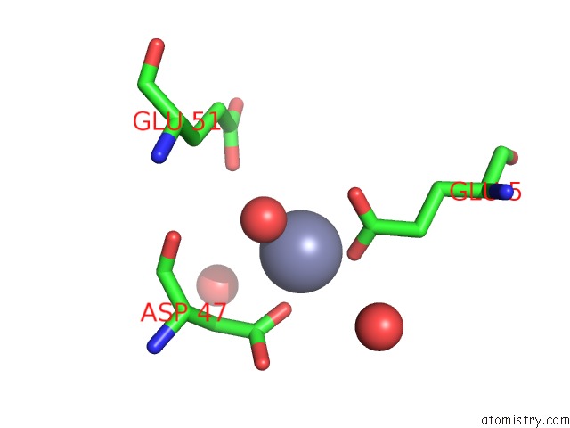
Mono view
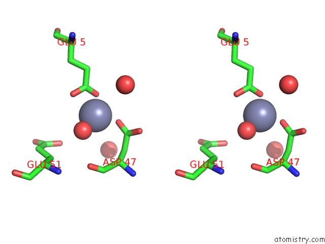
Stereo pair view
Mono view
Stereo pair view
A full contact list of Zinc with other atoms in the Zn binding
site number 1 of X-Ray Structure of Calcium-Loaded Calbindomodulin (A Calbindin D9K Re- Engineered to Undergo A Conformational Opening) at 1.44 A Resolution within 5.0Å range:
|
Zinc binding site 2 out of 2 in 1qx2
Go back to
Zinc binding site 2 out
of 2 in the X-Ray Structure of Calcium-Loaded Calbindomodulin (A Calbindin D9K Re- Engineered to Undergo A Conformational Opening) at 1.44 A Resolution
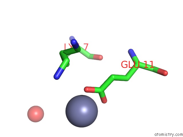
Mono view
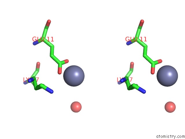
Stereo pair view
Mono view
Stereo pair view
A full contact list of Zinc with other atoms in the Zn binding
site number 2 of X-Ray Structure of Calcium-Loaded Calbindomodulin (A Calbindin D9K Re- Engineered to Undergo A Conformational Opening) at 1.44 A Resolution within 5.0Å range:
|
Reference:
C.G.Bunick,
M.R.Nelson,
S.Mangahas,
M.J.Hunter,
J.H.Sheehan,
L.S.Mizoue,
G.J.Bunick,
W.J.Chazin.
Designing Sequence to Control Protein Function in An Ef-Hand Protein J.Am.Chem.Soc. V. 126 5990 2004.
ISSN: ISSN 0002-7863
PubMed: 15137763
DOI: 10.1021/JA0397456
Page generated: Wed Oct 16 18:18:02 2024
ISSN: ISSN 0002-7863
PubMed: 15137763
DOI: 10.1021/JA0397456
Last articles
Zn in 9MJ5Zn in 9HNW
Zn in 9G0L
Zn in 9FNE
Zn in 9DZN
Zn in 9E0I
Zn in 9D32
Zn in 9DAK
Zn in 8ZXC
Zn in 8ZUF