Zinc »
PDB 1qmu-1r23 »
1qwu »
Zinc in PDB 1qwu: Golgi Alpha-Mannosidase II D341N Mutant Complex with 5-F-Guloside
Enzymatic activity of Golgi Alpha-Mannosidase II D341N Mutant Complex with 5-F-Guloside
All present enzymatic activity of Golgi Alpha-Mannosidase II D341N Mutant Complex with 5-F-Guloside:
3.2.1.114;
3.2.1.114;
Protein crystallography data
The structure of Golgi Alpha-Mannosidase II D341N Mutant Complex with 5-F-Guloside, PDB code: 1qwu
was solved by
S.Numao,
D.A.Kuntz,
S.G.Withers,
D.R.Rose,
with X-Ray Crystallography technique. A brief refinement statistics is given in the table below:
| Resolution Low / High (Å) | 19.84 / 2.03 |
| Space group | P 21 21 21 |
| Cell size a, b, c (Å), α, β, γ (°) | 68.850, 109.799, 138.765, 90.00, 90.00, 90.00 |
| R / Rfree (%) | 15.1 / 18.9 |
Other elements in 1qwu:
The structure of Golgi Alpha-Mannosidase II D341N Mutant Complex with 5-F-Guloside also contains other interesting chemical elements:
| Fluorine | (F) | 1 atom |
Zinc Binding Sites:
The binding sites of Zinc atom in the Golgi Alpha-Mannosidase II D341N Mutant Complex with 5-F-Guloside
(pdb code 1qwu). This binding sites where shown within
5.0 Angstroms radius around Zinc atom.
In total only one binding site of Zinc was determined in the Golgi Alpha-Mannosidase II D341N Mutant Complex with 5-F-Guloside, PDB code: 1qwu:
In total only one binding site of Zinc was determined in the Golgi Alpha-Mannosidase II D341N Mutant Complex with 5-F-Guloside, PDB code: 1qwu:
Zinc binding site 1 out of 1 in 1qwu
Go back to
Zinc binding site 1 out
of 1 in the Golgi Alpha-Mannosidase II D341N Mutant Complex with 5-F-Guloside
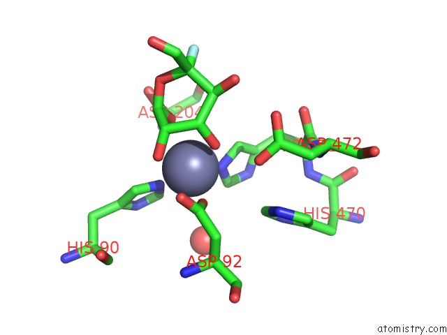
Mono view
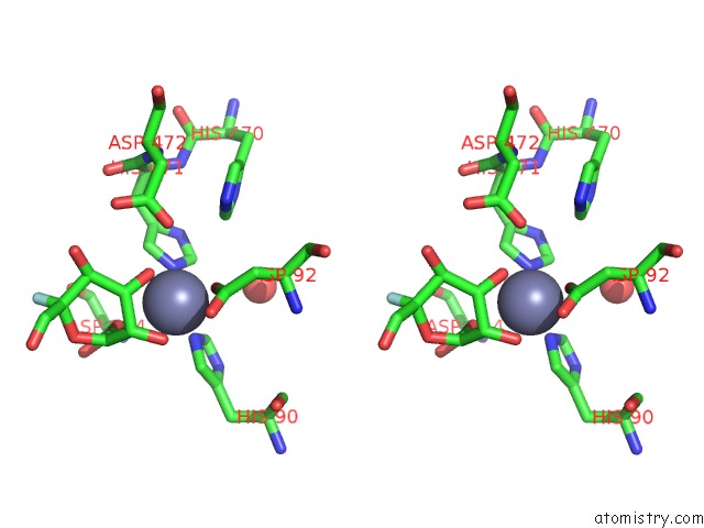
Stereo pair view
Mono view
Stereo pair view
A full contact list of Zinc with other atoms in the Zn binding
site number 1 of Golgi Alpha-Mannosidase II D341N Mutant Complex with 5-F-Guloside within 5.0Å range:
|
Reference:
S.Numao,
D.A.Kuntz,
S.G.Withers,
D.R.Rose.
Insights Into the Mechanism of Drosophila Melanogaster Golgi Alpha-Mannosidase II Through the Structural Analysis of Covalent Reaction Intermediates. J.Biol.Chem. V. 278 48074 2003.
ISSN: ISSN 0021-9258
PubMed: 12960159
DOI: 10.1074/JBC.M309249200
Page generated: Wed Oct 16 18:17:41 2024
ISSN: ISSN 0021-9258
PubMed: 12960159
DOI: 10.1074/JBC.M309249200
Last articles
Zn in 9MJ5Zn in 9HNW
Zn in 9G0L
Zn in 9FNE
Zn in 9DZN
Zn in 9E0I
Zn in 9D32
Zn in 9DAK
Zn in 8ZXC
Zn in 8ZUF