Zinc »
PDB 1q66-1qmd »
1q7l »
Zinc in PDB 1q7l: Zn-Binding Domain of the T347G Mutant of Human Aminoacylase- I
Enzymatic activity of Zn-Binding Domain of the T347G Mutant of Human Aminoacylase- I
All present enzymatic activity of Zn-Binding Domain of the T347G Mutant of Human Aminoacylase- I:
3.5.1.14;
3.5.1.14;
Protein crystallography data
The structure of Zn-Binding Domain of the T347G Mutant of Human Aminoacylase- I, PDB code: 1q7l
was solved by
H.A.Lindner,
V.V.Lunin,
A.Alary,
R.Hecker,
M.Cygler,
R.Menard,
with X-Ray Crystallography technique. A brief refinement statistics is given in the table below:
| Resolution Low / High (Å) | 33.71 / 1.40 |
| Space group | P 21 21 21 |
| Cell size a, b, c (Å), α, β, γ (°) | 53.528, 67.249, 146.479, 90.00, 90.00, 90.00 |
| R / Rfree (%) | 13.3 / 17.2 |
Zinc Binding Sites:
The binding sites of Zinc atom in the Zn-Binding Domain of the T347G Mutant of Human Aminoacylase- I
(pdb code 1q7l). This binding sites where shown within
5.0 Angstroms radius around Zinc atom.
In total 4 binding sites of Zinc where determined in the Zn-Binding Domain of the T347G Mutant of Human Aminoacylase- I, PDB code: 1q7l:
Jump to Zinc binding site number: 1; 2; 3; 4;
In total 4 binding sites of Zinc where determined in the Zn-Binding Domain of the T347G Mutant of Human Aminoacylase- I, PDB code: 1q7l:
Jump to Zinc binding site number: 1; 2; 3; 4;
Zinc binding site 1 out of 4 in 1q7l
Go back to
Zinc binding site 1 out
of 4 in the Zn-Binding Domain of the T347G Mutant of Human Aminoacylase- I
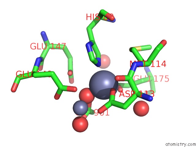
Mono view

Stereo pair view
Mono view
Stereo pair view
A full contact list of Zinc with other atoms in the Zn binding
site number 1 of Zn-Binding Domain of the T347G Mutant of Human Aminoacylase- I within 5.0Å range:
|
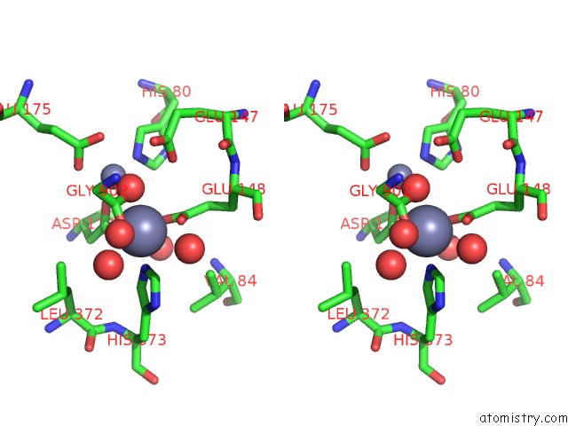
Zinc binding site 2 out of 4 in 1q7l
Go back to
Zinc binding site 2 out
of 4 in the Zn-Binding Domain of the T347G Mutant of Human Aminoacylase- I

Mono view

Stereo pair view
Mono view
Stereo pair view
A full contact list of Zinc with other atoms in the Zn binding
site number 2 of Zn-Binding Domain of the T347G Mutant of Human Aminoacylase- I within 5.0Å range:
|
Zinc binding site 3 out of 4 in 1q7l
Go back to
Zinc binding site 3 out
of 4 in the Zn-Binding Domain of the T347G Mutant of Human Aminoacylase- I

Mono view

Stereo pair view
Mono view
Stereo pair view
A full contact list of Zinc with other atoms in the Zn binding
site number 3 of Zn-Binding Domain of the T347G Mutant of Human Aminoacylase- I within 5.0Å range:
|
Zinc binding site 4 out of 4 in 1q7l
Go back to
Zinc binding site 4 out
of 4 in the Zn-Binding Domain of the T347G Mutant of Human Aminoacylase- I

Mono view
Stereo pair view
Mono view
Stereo pair view
A full contact list of Zinc with other atoms in the Zn binding
site number 4 of Zn-Binding Domain of the T347G Mutant of Human Aminoacylase- I within 5.0Å range:
|
Reference:
H.A.Lindner,
V.V.Lunin,
A.Alary,
R.Hecker,
M.Cygler,
R.Menard.
Essential Roles of Zinc Ligation and Enzyme Dimerization For Catalysis in the Aminoacylase-1/M20 Family. J.Biol.Chem. V. 278 44496 2003.
ISSN: ISSN 0021-9258
PubMed: 12933810
DOI: 10.1074/JBC.M304233200
Page generated: Wed Oct 16 18:05:44 2024
ISSN: ISSN 0021-9258
PubMed: 12933810
DOI: 10.1074/JBC.M304233200
Last articles
Zn in 9MJ5Zn in 9HNW
Zn in 9G0L
Zn in 9FNE
Zn in 9DZN
Zn in 9E0I
Zn in 9D32
Zn in 9DAK
Zn in 8ZXC
Zn in 8ZUF