Zinc »
PDB 1p1v-1ped »
1p6n »
Zinc in PDB 1p6n: Bovine Endothelial Nos Heme Domain with L-N(Omega)-Nitroarginine-(4R)- Amino-L-Proline Amide Bound
Enzymatic activity of Bovine Endothelial Nos Heme Domain with L-N(Omega)-Nitroarginine-(4R)- Amino-L-Proline Amide Bound
All present enzymatic activity of Bovine Endothelial Nos Heme Domain with L-N(Omega)-Nitroarginine-(4R)- Amino-L-Proline Amide Bound:
1.14.13.39;
1.14.13.39;
Protein crystallography data
The structure of Bovine Endothelial Nos Heme Domain with L-N(Omega)-Nitroarginine-(4R)- Amino-L-Proline Amide Bound, PDB code: 1p6n
was solved by
M.L.Flinspach,
H.Li,
J.Jamal,
W.Yang,
H.Huang,
J.-M.Hah,
J.A.Gomez-Vidal,
E.A.Litzinger,
R.B.Silverman,
T.L.Poulos,
with X-Ray Crystallography technique. A brief refinement statistics is given in the table below:
| Resolution Low / High (Å) | 42.55 / 2.50 |
| Space group | P 21 21 21 |
| Cell size a, b, c (Å), α, β, γ (°) | 57.669, 106.292, 156.524, 90.00, 90.00, 90.00 |
| R / Rfree (%) | 21.8 / 27.8 |
Other elements in 1p6n:
The structure of Bovine Endothelial Nos Heme Domain with L-N(Omega)-Nitroarginine-(4R)- Amino-L-Proline Amide Bound also contains other interesting chemical elements:
| Arsenic | (As) | 2 atoms |
| Iron | (Fe) | 2 atoms |
Zinc Binding Sites:
The binding sites of Zinc atom in the Bovine Endothelial Nos Heme Domain with L-N(Omega)-Nitroarginine-(4R)- Amino-L-Proline Amide Bound
(pdb code 1p6n). This binding sites where shown within
5.0 Angstroms radius around Zinc atom.
In total only one binding site of Zinc was determined in the Bovine Endothelial Nos Heme Domain with L-N(Omega)-Nitroarginine-(4R)- Amino-L-Proline Amide Bound, PDB code: 1p6n:
In total only one binding site of Zinc was determined in the Bovine Endothelial Nos Heme Domain with L-N(Omega)-Nitroarginine-(4R)- Amino-L-Proline Amide Bound, PDB code: 1p6n:
Zinc binding site 1 out of 1 in 1p6n
Go back to
Zinc binding site 1 out
of 1 in the Bovine Endothelial Nos Heme Domain with L-N(Omega)-Nitroarginine-(4R)- Amino-L-Proline Amide Bound
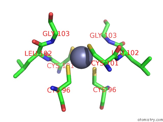
Mono view
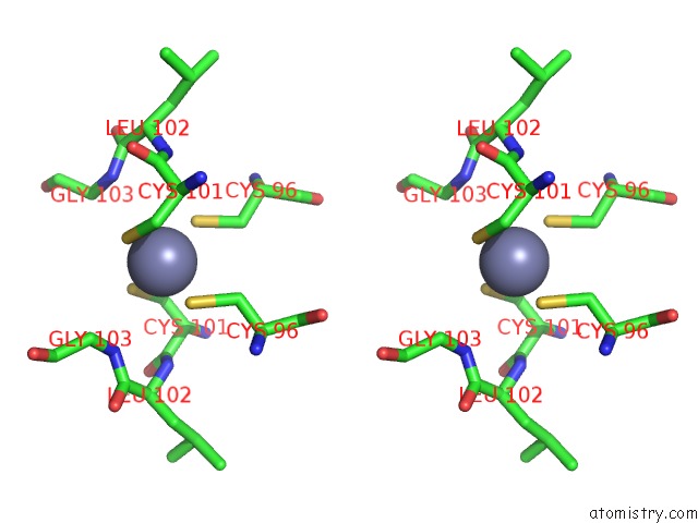
Stereo pair view
Mono view
Stereo pair view
A full contact list of Zinc with other atoms in the Zn binding
site number 1 of Bovine Endothelial Nos Heme Domain with L-N(Omega)-Nitroarginine-(4R)- Amino-L-Proline Amide Bound within 5.0Å range:
|
Reference:
M.L.Flinspach,
H.Li,
J.Jamal,
W.Yang,
H.Huang,
J.M.Hah,
J.A.Gomez-Vidal,
E.A.Litzinger,
R.B.Silverman,
T.L.Poulos.
Structural Basis For Dipeptide Amide Isoform-Selective Inhibition of Neuronal Nitric Oxide Synthase. Nat.Struct.Mol.Biol. V. 11 54 2004.
ISSN: ISSN 1545-9993
PubMed: 14718923
DOI: 10.1038/NSMB704
Page generated: Wed Oct 16 17:45:34 2024
ISSN: ISSN 1545-9993
PubMed: 14718923
DOI: 10.1038/NSMB704
Last articles
Zn in 9MJ5Zn in 9HNW
Zn in 9G0L
Zn in 9FNE
Zn in 9DZN
Zn in 9E0I
Zn in 9D32
Zn in 9DAK
Zn in 8ZXC
Zn in 8ZUF