Zinc »
PDB 1ml9-1my1 »
1mr1 »
Zinc in PDB 1mr1: Crystal Structure of A SMAD4-Ski Complex
Protein crystallography data
The structure of Crystal Structure of A SMAD4-Ski Complex, PDB code: 1mr1
was solved by
J.-W.Wu,
A.R.Krawitz,
J.Chai,
W.Li,
F.Zhang,
K.Luo,
Y.Shi,
with X-Ray Crystallography technique. A brief refinement statistics is given in the table below:
| Resolution Low / High (Å) | 20.00 / 2.85 |
| Space group | P 32 2 1 |
| Cell size a, b, c (Å), α, β, γ (°) | 109.800, 109.800, 141.100, 90.00, 90.00, 120.00 |
| R / Rfree (%) | 23.1 / 28 |
Zinc Binding Sites:
The binding sites of Zinc atom in the Crystal Structure of A SMAD4-Ski Complex
(pdb code 1mr1). This binding sites where shown within
5.0 Angstroms radius around Zinc atom.
In total 2 binding sites of Zinc where determined in the Crystal Structure of A SMAD4-Ski Complex, PDB code: 1mr1:
Jump to Zinc binding site number: 1; 2;
In total 2 binding sites of Zinc where determined in the Crystal Structure of A SMAD4-Ski Complex, PDB code: 1mr1:
Jump to Zinc binding site number: 1; 2;
Zinc binding site 1 out of 2 in 1mr1
Go back to
Zinc binding site 1 out
of 2 in the Crystal Structure of A SMAD4-Ski Complex
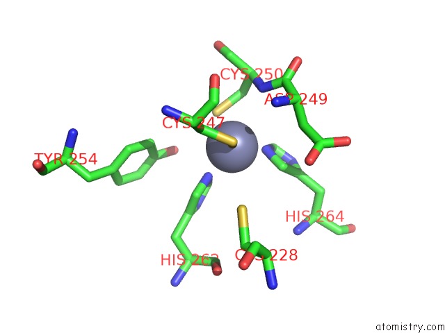
Mono view
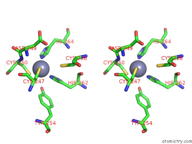
Stereo pair view
Mono view
Stereo pair view
A full contact list of Zinc with other atoms in the Zn binding
site number 1 of Crystal Structure of A SMAD4-Ski Complex within 5.0Å range:
|
Zinc binding site 2 out of 2 in 1mr1
Go back to
Zinc binding site 2 out
of 2 in the Crystal Structure of A SMAD4-Ski Complex
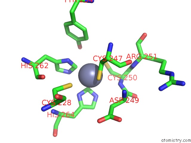
Mono view
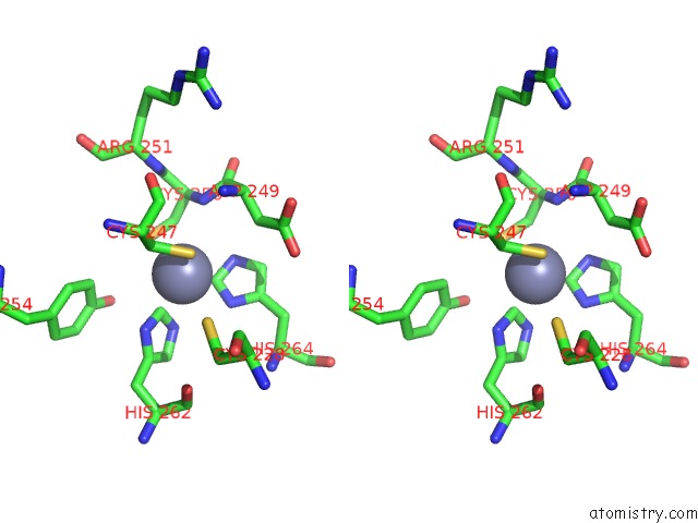
Stereo pair view
Mono view
Stereo pair view
A full contact list of Zinc with other atoms in the Zn binding
site number 2 of Crystal Structure of A SMAD4-Ski Complex within 5.0Å range:
|
Reference:
J.-W.Wu,
A.R.Krawitz,
J.Chai,
W.Li,
F.Zhang,
K.Luo,
Y.Shi.
Structural Mechanism of SMAD4 Recognition By the Nuclear Oncoprotein Ski: Insights on Ski-Mediated Repression of Tgf-Beta Signaling Cell(Cambridge,Mass.) V. 111 357 2002.
ISSN: ISSN 0092-8674
PubMed: 12419246
DOI: 10.1016/S0092-8674(02)01006-1
Page generated: Wed Oct 16 17:01:39 2024
ISSN: ISSN 0092-8674
PubMed: 12419246
DOI: 10.1016/S0092-8674(02)01006-1
Last articles
Zn in 9MJ5Zn in 9HNW
Zn in 9G0L
Zn in 9FNE
Zn in 9DZN
Zn in 9E0I
Zn in 9D32
Zn in 9DAK
Zn in 8ZXC
Zn in 8ZUF