Zinc »
PDB 1lgd-1m2n »
1lml »
Zinc in PDB 1lml: Leishmanolysin
Enzymatic activity of Leishmanolysin
All present enzymatic activity of Leishmanolysin:
3.4.24.36;
3.4.24.36;
Protein crystallography data
The structure of Leishmanolysin, PDB code: 1lml
was solved by
E.Schlagenhauf,
R.Etges,
P.Metcalf,
with X-Ray Crystallography technique. A brief refinement statistics is given in the table below:
| Resolution Low / High (Å) | 5.00 / 1.86 |
| Space group | C 1 2 1 |
| Cell size a, b, c (Å), α, β, γ (°) | 106.325, 90.144, 70.145, 90.00, 110.54, 90.00 |
| R / Rfree (%) | 19.1 / 20.8 |
Zinc Binding Sites:
The binding sites of Zinc atom in the Leishmanolysin
(pdb code 1lml). This binding sites where shown within
5.0 Angstroms radius around Zinc atom.
In total only one binding site of Zinc was determined in the Leishmanolysin, PDB code: 1lml:
In total only one binding site of Zinc was determined in the Leishmanolysin, PDB code: 1lml:
Zinc binding site 1 out of 1 in 1lml
Go back to
Zinc binding site 1 out
of 1 in the Leishmanolysin
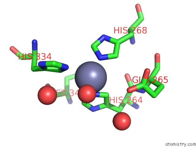
Mono view
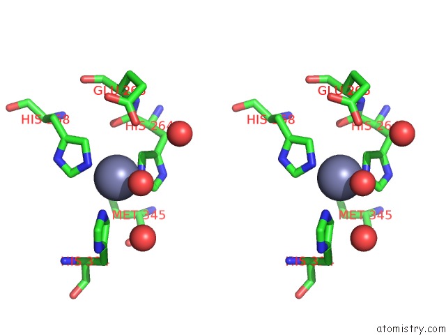
Stereo pair view
Mono view
Stereo pair view
A full contact list of Zinc with other atoms in the Zn binding
site number 1 of Leishmanolysin within 5.0Å range:
|
Reference:
E.Schlagenhauf,
R.Etges,
P.Metcalf.
The Crystal Structure of the Leishmania Major Surface Proteinase Leishmanolysin (GP63). Structure V. 6 1035 1998.
ISSN: ISSN 0969-2126
PubMed: 9739094
DOI: 10.1016/S0969-2126(98)00104-X
Page generated: Tue Aug 19 21:34:14 2025
ISSN: ISSN 0969-2126
PubMed: 9739094
DOI: 10.1016/S0969-2126(98)00104-X
Last articles
Zn in 1WIRZn in 1WIL
Zn in 1WII
Zn in 1WG2
Zn in 1WIG
Zn in 1WGE
Zn in 1WFZ
Zn in 1WFP
Zn in 1WFL
Zn in 1WFX