Zinc »
PDB 1kk1-1kzp »
1kl9 »
Zinc in PDB 1kl9: Crystal Structure of the N-Terminal Segment of Human Eukaryotic Initiation Factor 2ALPHA
Protein crystallography data
The structure of Crystal Structure of the N-Terminal Segment of Human Eukaryotic Initiation Factor 2ALPHA, PDB code: 1kl9
was solved by
M.C.Nonato,
J.Widom,
J.Clardy,
with X-Ray Crystallography technique. A brief refinement statistics is given in the table below:
| Resolution Low / High (Å) | 30.00 / 1.90 |
| Space group | P 2 2 21 |
| Cell size a, b, c (Å), α, β, γ (°) | 37.280, 44.200, 121.420, 90.00, 90.00, 90.00 |
| R / Rfree (%) | 19.8 / 23.3 |
Zinc Binding Sites:
The binding sites of Zinc atom in the Crystal Structure of the N-Terminal Segment of Human Eukaryotic Initiation Factor 2ALPHA
(pdb code 1kl9). This binding sites where shown within
5.0 Angstroms radius around Zinc atom.
In total 4 binding sites of Zinc where determined in the Crystal Structure of the N-Terminal Segment of Human Eukaryotic Initiation Factor 2ALPHA, PDB code: 1kl9:
Jump to Zinc binding site number: 1; 2; 3; 4;
In total 4 binding sites of Zinc where determined in the Crystal Structure of the N-Terminal Segment of Human Eukaryotic Initiation Factor 2ALPHA, PDB code: 1kl9:
Jump to Zinc binding site number: 1; 2; 3; 4;
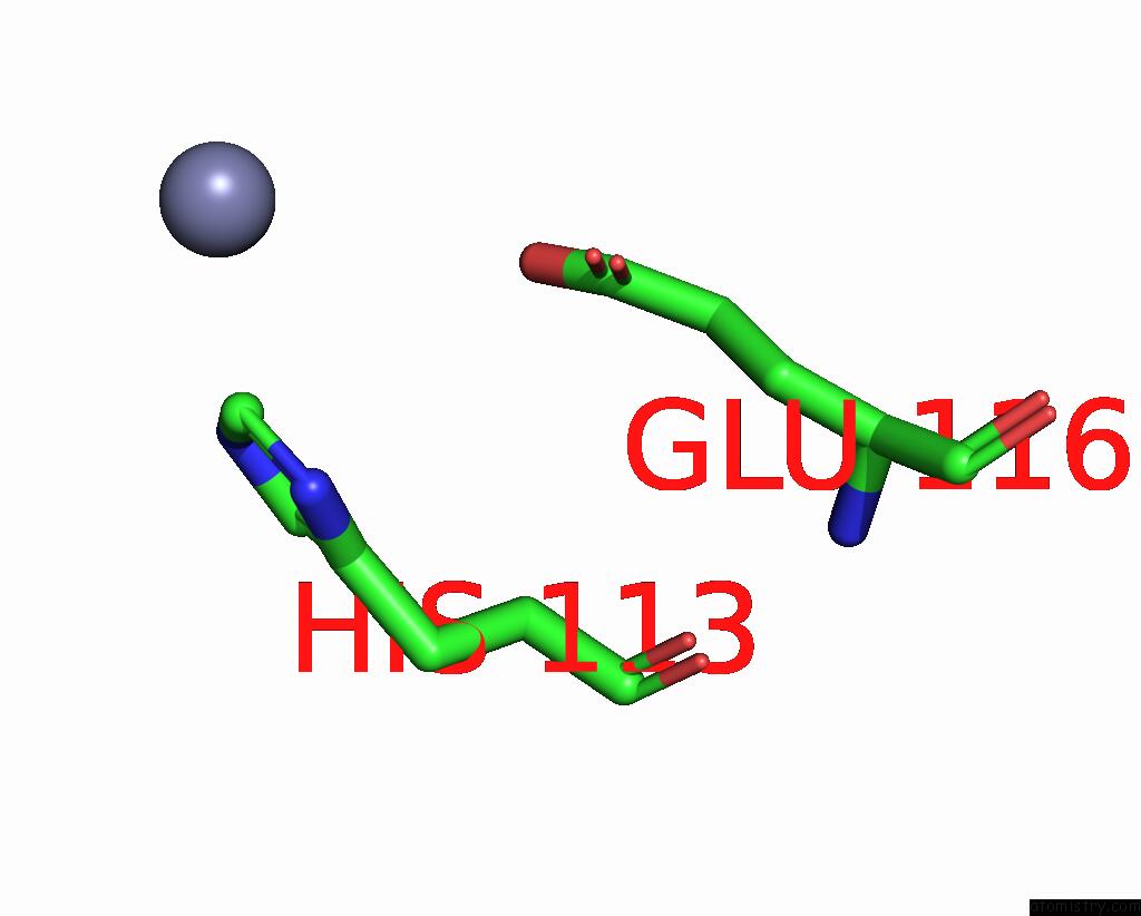
Zinc binding site 1 out of 4 in 1kl9
Go back to
Zinc binding site 1 out
of 4 in the Crystal Structure of the N-Terminal Segment of Human Eukaryotic Initiation Factor 2ALPHA

Mono view

Stereo pair view
Mono view
Stereo pair view
A full contact list of Zinc with other atoms in the Zn binding
site number 1 of Crystal Structure of the N-Terminal Segment of Human Eukaryotic Initiation Factor 2ALPHA within 5.0Å range:
|
Zinc binding site 2 out of 4 in 1kl9
Go back to
Zinc binding site 2 out
of 4 in the Crystal Structure of the N-Terminal Segment of Human Eukaryotic Initiation Factor 2ALPHA

Mono view

Stereo pair view
Mono view
Stereo pair view
A full contact list of Zinc with other atoms in the Zn binding
site number 2 of Crystal Structure of the N-Terminal Segment of Human Eukaryotic Initiation Factor 2ALPHA within 5.0Å range:
|
Zinc binding site 3 out of 4 in 1kl9
Go back to
Zinc binding site 3 out
of 4 in the Crystal Structure of the N-Terminal Segment of Human Eukaryotic Initiation Factor 2ALPHA

Mono view

Stereo pair view
Mono view
Stereo pair view
A full contact list of Zinc with other atoms in the Zn binding
site number 3 of Crystal Structure of the N-Terminal Segment of Human Eukaryotic Initiation Factor 2ALPHA within 5.0Å range:
|
Zinc binding site 4 out of 4 in 1kl9
Go back to
Zinc binding site 4 out
of 4 in the Crystal Structure of the N-Terminal Segment of Human Eukaryotic Initiation Factor 2ALPHA
Mono view

Stereo pair view
Mono view
Stereo pair view
A full contact list of Zinc with other atoms in the Zn binding
site number 4 of Crystal Structure of the N-Terminal Segment of Human Eukaryotic Initiation Factor 2ALPHA within 5.0Å range:
|
Reference:
M.C.Nonato,
J.Widom,
J.Clardy.
Crystal Structure of the N-Terminal Segment of Human Eukaryotic Translation Initiation Factor 2ALPHA J.Biol.Chem. V. 277 17057 2002.
ISSN: ISSN 0021-9258
PubMed: 11859078
DOI: 10.1074/JBC.M111804200
Page generated: Sun Oct 13 04:31:08 2024
ISSN: ISSN 0021-9258
PubMed: 11859078
DOI: 10.1074/JBC.M111804200
Last articles
Zn in 9MJ5Zn in 9HNW
Zn in 9G0L
Zn in 9FNE
Zn in 9DZN
Zn in 9E0I
Zn in 9D32
Zn in 9DAK
Zn in 8ZXC
Zn in 8ZUF