Zinc »
PDB 1jpu-1k53 »
1jwq »
Zinc in PDB 1jwq: Structure of the Catalytic Domain of Cwlv, N-Acetylmuramoyl- L-Alanine Amidase From Bacillus(Paenibacillus) Polymyxa Var.Colistinus
Enzymatic activity of Structure of the Catalytic Domain of Cwlv, N-Acetylmuramoyl- L-Alanine Amidase From Bacillus(Paenibacillus) Polymyxa Var.Colistinus
All present enzymatic activity of Structure of the Catalytic Domain of Cwlv, N-Acetylmuramoyl- L-Alanine Amidase From Bacillus(Paenibacillus) Polymyxa Var.Colistinus:
3.5.1.28;
3.5.1.28;
Protein crystallography data
The structure of Structure of the Catalytic Domain of Cwlv, N-Acetylmuramoyl- L-Alanine Amidase From Bacillus(Paenibacillus) Polymyxa Var.Colistinus, PDB code: 1jwq
was solved by
T.Yamane,
Y.Koyama,
Y.Nojiri,
T.Hikage,
M.Akita,
A.Suzuki,
T.Shirai,
F.Ise,
T.Shida,
J.Sekiguchi,
with X-Ray Crystallography technique. A brief refinement statistics is given in the table below:
| Resolution Low / High (Å) | 20.00 / 1.80 |
| Space group | P 61 |
| Cell size a, b, c (Å), α, β, γ (°) | 66.500, 66.500, 88.340, 90.00, 90.00, 120.00 |
| R / Rfree (%) | 17.6 / 20.6 |
Zinc Binding Sites:
The binding sites of Zinc atom in the Structure of the Catalytic Domain of Cwlv, N-Acetylmuramoyl- L-Alanine Amidase From Bacillus(Paenibacillus) Polymyxa Var.Colistinus
(pdb code 1jwq). This binding sites where shown within
5.0 Angstroms radius around Zinc atom.
In total only one binding site of Zinc was determined in the Structure of the Catalytic Domain of Cwlv, N-Acetylmuramoyl- L-Alanine Amidase From Bacillus(Paenibacillus) Polymyxa Var.Colistinus, PDB code: 1jwq:
In total only one binding site of Zinc was determined in the Structure of the Catalytic Domain of Cwlv, N-Acetylmuramoyl- L-Alanine Amidase From Bacillus(Paenibacillus) Polymyxa Var.Colistinus, PDB code: 1jwq:
Zinc binding site 1 out of 1 in 1jwq
Go back to
Zinc binding site 1 out
of 1 in the Structure of the Catalytic Domain of Cwlv, N-Acetylmuramoyl- L-Alanine Amidase From Bacillus(Paenibacillus) Polymyxa Var.Colistinus

Mono view
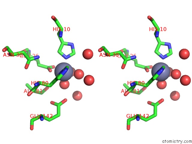
Stereo pair view
Mono view
Stereo pair view
A full contact list of Zinc with other atoms in the Zn binding
site number 1 of Structure of the Catalytic Domain of Cwlv, N-Acetylmuramoyl- L-Alanine Amidase From Bacillus(Paenibacillus) Polymyxa Var.Colistinus within 5.0Å range:
|
Reference:
T.Yamane,
Y.Koyama,
Y.Nojiri,
T.Hikage,
M.Akita,
A.Suzuki,
T.Shirai,
F.Ise,
T.Shida,
J.Sekiguchi.
The Structure of the Catalytic Domain of N-Acetylmuramoyl-L-Alanine Amidase, A Cell Wall Hydrolase From Bacillus Polymyxa Var.Colistinus and Its Resemblance to the Structure of Carboxypeptidases To Be Published.
Page generated: Sun Oct 13 03:54:32 2024
Last articles
Zn in 9MJ5Zn in 9HNW
Zn in 9G0L
Zn in 9FNE
Zn in 9DZN
Zn in 9E0I
Zn in 9D32
Zn in 9DAK
Zn in 8ZXC
Zn in 8ZUF