Zinc »
PDB 1jao-1joe »
1jcr »
Zinc in PDB 1jcr: Crystal Structure of Rat Protein Farnesyltransferase Complexed with the Non-Substrate Tetrapeptide Inhibitor Cvfm and Farnesyl Diphosphate Substrate
Protein crystallography data
The structure of Crystal Structure of Rat Protein Farnesyltransferase Complexed with the Non-Substrate Tetrapeptide Inhibitor Cvfm and Farnesyl Diphosphate Substrate, PDB code: 1jcr
was solved by
S.B.Long,
P.J.Casey,
L.S.Beese,
with X-Ray Crystallography technique. A brief refinement statistics is given in the table below:
| Resolution Low / High (Å) | 50.00 / 2.00 |
| Space group | P 61 |
| Cell size a, b, c (Å), α, β, γ (°) | 171.225, 171.225, 69.332, 90.00, 90.00, 120.00 |
| R / Rfree (%) | 16.6 / 20.6 |
Zinc Binding Sites:
The binding sites of Zinc atom in the Crystal Structure of Rat Protein Farnesyltransferase Complexed with the Non-Substrate Tetrapeptide Inhibitor Cvfm and Farnesyl Diphosphate Substrate
(pdb code 1jcr). This binding sites where shown within
5.0 Angstroms radius around Zinc atom.
In total only one binding site of Zinc was determined in the Crystal Structure of Rat Protein Farnesyltransferase Complexed with the Non-Substrate Tetrapeptide Inhibitor Cvfm and Farnesyl Diphosphate Substrate, PDB code: 1jcr:
In total only one binding site of Zinc was determined in the Crystal Structure of Rat Protein Farnesyltransferase Complexed with the Non-Substrate Tetrapeptide Inhibitor Cvfm and Farnesyl Diphosphate Substrate, PDB code: 1jcr:
Zinc binding site 1 out of 1 in 1jcr
Go back to
Zinc binding site 1 out
of 1 in the Crystal Structure of Rat Protein Farnesyltransferase Complexed with the Non-Substrate Tetrapeptide Inhibitor Cvfm and Farnesyl Diphosphate Substrate

Mono view
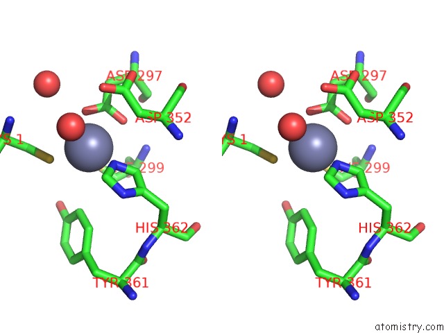
Stereo pair view
Mono view
Stereo pair view
A full contact list of Zinc with other atoms in the Zn binding
site number 1 of Crystal Structure of Rat Protein Farnesyltransferase Complexed with the Non-Substrate Tetrapeptide Inhibitor Cvfm and Farnesyl Diphosphate Substrate within 5.0Å range:
|
Reference:
S.B.Long,
P.J.Hancock,
A.M.Kral,
H.W.Hellinga,
L.S.Beese.
The Crystal Structure of Human Protein Farnesyltransferase Reveals the Basis For Inhibition By Caax Tetrapeptides and Their Mimetics. Proc.Natl.Acad.Sci.Usa V. 98 12948 2001.
ISSN: ISSN 0027-8424
PubMed: 11687658
DOI: 10.1073/PNAS.241407898
Page generated: Sun Oct 13 03:30:21 2024
ISSN: ISSN 0027-8424
PubMed: 11687658
DOI: 10.1073/PNAS.241407898
Last articles
Zn in 9MJ5Zn in 9HNW
Zn in 9G0L
Zn in 9FNE
Zn in 9DZN
Zn in 9E0I
Zn in 9D32
Zn in 9DAK
Zn in 8ZXC
Zn in 8ZUF