Zinc »
PDB 1i96-1im5 »
1ib5 »
Zinc in PDB 1ib5: X-Ray 3D Structure of P.Leiognathi Cu,Zn Sod Mutant W83Y
Enzymatic activity of X-Ray 3D Structure of P.Leiognathi Cu,Zn Sod Mutant W83Y
All present enzymatic activity of X-Ray 3D Structure of P.Leiognathi Cu,Zn Sod Mutant W83Y:
1.15.1.1;
1.15.1.1;
Protein crystallography data
The structure of X-Ray 3D Structure of P.Leiognathi Cu,Zn Sod Mutant W83Y, PDB code: 1ib5
was solved by
M.E.Stroppolo,
A.Pesce,
M.D'orazio,
P.O'neill,
D.Bordo,
C.Rosano,
M.Milani,
A.Battistoni,
M.Bolognesi,
A.Desideri,
with X-Ray Crystallography technique. A brief refinement statistics is given in the table below:
| Resolution Low / High (Å) | 20.00 / 2.45 |
| Space group | H 3 2 |
| Cell size a, b, c (Å), α, β, γ (°) | 87.336, 87.336, 98.223, 90.00, 90.00, 120.00 |
| R / Rfree (%) | 21.6 / 26.5 |
Other elements in 1ib5:
The structure of X-Ray 3D Structure of P.Leiognathi Cu,Zn Sod Mutant W83Y also contains other interesting chemical elements:
| Copper | (Cu) | 1 atom |
Zinc Binding Sites:
The binding sites of Zinc atom in the X-Ray 3D Structure of P.Leiognathi Cu,Zn Sod Mutant W83Y
(pdb code 1ib5). This binding sites where shown within
5.0 Angstroms radius around Zinc atom.
In total only one binding site of Zinc was determined in the X-Ray 3D Structure of P.Leiognathi Cu,Zn Sod Mutant W83Y, PDB code: 1ib5:
In total only one binding site of Zinc was determined in the X-Ray 3D Structure of P.Leiognathi Cu,Zn Sod Mutant W83Y, PDB code: 1ib5:
Zinc binding site 1 out of 1 in 1ib5
Go back to
Zinc binding site 1 out
of 1 in the X-Ray 3D Structure of P.Leiognathi Cu,Zn Sod Mutant W83Y
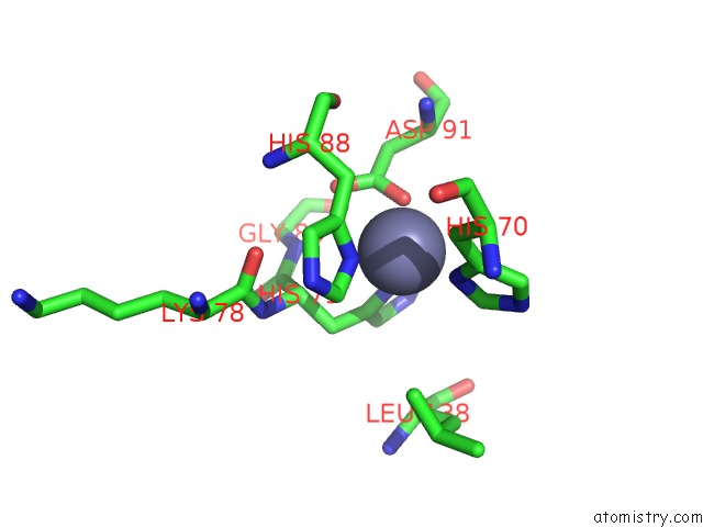
Mono view
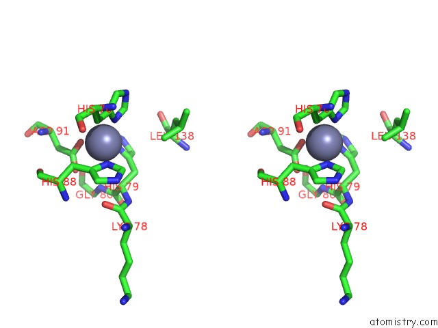
Stereo pair view
Mono view
Stereo pair view
A full contact list of Zinc with other atoms in the Zn binding
site number 1 of X-Ray 3D Structure of P.Leiognathi Cu,Zn Sod Mutant W83Y within 5.0Å range:
|
Reference:
M.E.Stroppolo,
A.Pesce,
M.D'orazio,
P.O'neill,
D.Bordo,
C.Rosano,
M.Milani,
A.Battistoni,
M.Bolognesi,
A.Desideri.
Single Mutations at the Subunit Interface Modulate Copper Reactivity in Photobacterium Leiognathi Cu,Zn Superoxide Dismutase. J.Mol.Biol. V. 308 555 2001.
ISSN: ISSN 0022-2836
PubMed: 11327787
DOI: 10.1006/JMBI.2001.4606
Page generated: Sun Oct 13 03:02:00 2024
ISSN: ISSN 0022-2836
PubMed: 11327787
DOI: 10.1006/JMBI.2001.4606
Last articles
Zn in 9MJ5Zn in 9HNW
Zn in 9G0L
Zn in 9FNE
Zn in 9DZN
Zn in 9E0I
Zn in 9D32
Zn in 9DAK
Zn in 8ZXC
Zn in 8ZUF