Zinc »
PDB 1hxy-1i95 »
1i5o »
Zinc in PDB 1i5o: Crystal Structure of Mutant R105A of E. Coli Aspartate Transcarbamoylase
Enzymatic activity of Crystal Structure of Mutant R105A of E. Coli Aspartate Transcarbamoylase
All present enzymatic activity of Crystal Structure of Mutant R105A of E. Coli Aspartate Transcarbamoylase:
2.1.3.2;
2.1.3.2;
Protein crystallography data
The structure of Crystal Structure of Mutant R105A of E. Coli Aspartate Transcarbamoylase, PDB code: 1i5o
was solved by
C.P.Macol,
H.Tsuruta,
B.Stec,
E.R.Kantrowitz,
with X-Ray Crystallography technique. A brief refinement statistics is given in the table below:
| Resolution Low / High (Å) | 10.00 / 2.80 |
| Space group | P 3 2 1 |
| Cell size a, b, c (Å), α, β, γ (°) | 122.250, 122.250, 142.670, 90.00, 90.00, 120.00 |
| R / Rfree (%) | 15.5 / 21.2 |
Zinc Binding Sites:
The binding sites of Zinc atom in the Crystal Structure of Mutant R105A of E. Coli Aspartate Transcarbamoylase
(pdb code 1i5o). This binding sites where shown within
5.0 Angstroms radius around Zinc atom.
In total 2 binding sites of Zinc where determined in the Crystal Structure of Mutant R105A of E. Coli Aspartate Transcarbamoylase, PDB code: 1i5o:
Jump to Zinc binding site number: 1; 2;
In total 2 binding sites of Zinc where determined in the Crystal Structure of Mutant R105A of E. Coli Aspartate Transcarbamoylase, PDB code: 1i5o:
Jump to Zinc binding site number: 1; 2;
Zinc binding site 1 out of 2 in 1i5o
Go back to
Zinc binding site 1 out
of 2 in the Crystal Structure of Mutant R105A of E. Coli Aspartate Transcarbamoylase
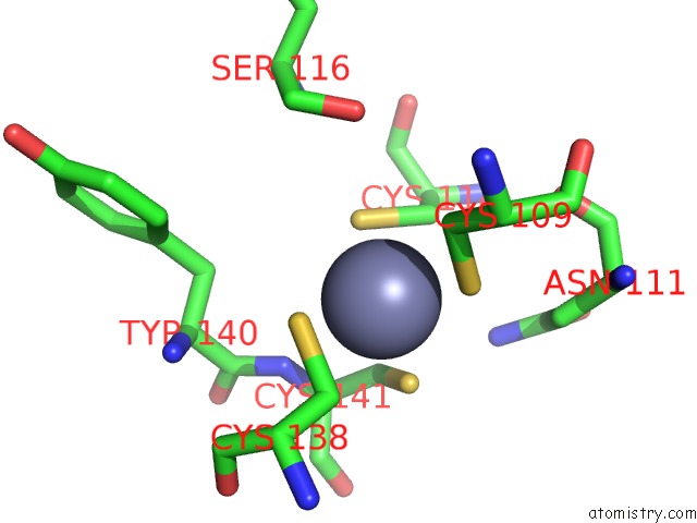
Mono view

Stereo pair view
Mono view
Stereo pair view
A full contact list of Zinc with other atoms in the Zn binding
site number 1 of Crystal Structure of Mutant R105A of E. Coli Aspartate Transcarbamoylase within 5.0Å range:
|
Zinc binding site 2 out of 2 in 1i5o
Go back to
Zinc binding site 2 out
of 2 in the Crystal Structure of Mutant R105A of E. Coli Aspartate Transcarbamoylase
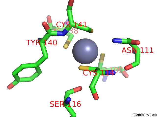
Mono view
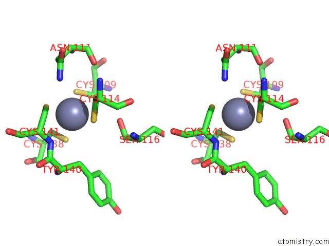
Stereo pair view
Mono view
Stereo pair view
A full contact list of Zinc with other atoms in the Zn binding
site number 2 of Crystal Structure of Mutant R105A of E. Coli Aspartate Transcarbamoylase within 5.0Å range:
|
Reference:
C.P.Macol,
H.Tsuruta,
B.Stec,
E.R.Kantrowitz.
Direct Structural Evidence For A Concerted Allosteric Transition in Escherichia Coli Aspartate Transcarbamoylase. Nat.Struct.Biol. V. 8 423 2001.
ISSN: ISSN 1072-8368
PubMed: 11323717
DOI: 10.1038/87582
Page generated: Sun Oct 13 02:52:00 2024
ISSN: ISSN 1072-8368
PubMed: 11323717
DOI: 10.1038/87582
Last articles
Zn in 9MJ5Zn in 9HNW
Zn in 9G0L
Zn in 9FNE
Zn in 9DZN
Zn in 9E0I
Zn in 9D32
Zn in 9DAK
Zn in 8ZXC
Zn in 8ZUF