Zinc »
PDB 1hxy-1i95 »
1i4p »
Zinc in PDB 1i4p: Crystal Structure of Staphylococcal Enterotoxin C2 at 100K Crystallized at pH 5.5
Protein crystallography data
The structure of Crystal Structure of Staphylococcal Enterotoxin C2 at 100K Crystallized at pH 5.5, PDB code: 1i4p
was solved by
D.Kumaran,
S.Swaminathan,
with X-Ray Crystallography technique. A brief refinement statistics is given in the table below:
| Resolution Low / High (Å) | 50.00 / 2.00 |
| Space group | P 43 21 2 |
| Cell size a, b, c (Å), α, β, γ (°) | 43.160, 43.160, 291.280, 90.00, 90.00, 90.00 |
| R / Rfree (%) | 20.6 / 26.3 |
Zinc Binding Sites:
The binding sites of Zinc atom in the Crystal Structure of Staphylococcal Enterotoxin C2 at 100K Crystallized at pH 5.5
(pdb code 1i4p). This binding sites where shown within
5.0 Angstroms radius around Zinc atom.
In total only one binding site of Zinc was determined in the Crystal Structure of Staphylococcal Enterotoxin C2 at 100K Crystallized at pH 5.5, PDB code: 1i4p:
In total only one binding site of Zinc was determined in the Crystal Structure of Staphylococcal Enterotoxin C2 at 100K Crystallized at pH 5.5, PDB code: 1i4p:
Zinc binding site 1 out of 1 in 1i4p
Go back to
Zinc binding site 1 out
of 1 in the Crystal Structure of Staphylococcal Enterotoxin C2 at 100K Crystallized at pH 5.5
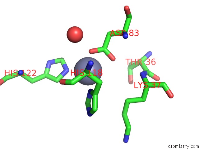
Mono view
Stereo pair view
Mono view
Stereo pair view
A full contact list of Zinc with other atoms in the Zn binding
site number 1 of Crystal Structure of Staphylococcal Enterotoxin C2 at 100K Crystallized at pH 5.5 within 5.0Å range:
|
Reference:
D.Kumaran,
S.Eswaramoorthy,
W.Furey,
M.Sax,
S.Swaminathan.
Structure of Staphylococcal Enterotoxin C2 at Various pH Levels. Acta Crystallogr.,Sect.D V. 57 1270 2001.
ISSN: ISSN 0907-4449
PubMed: 11526318
DOI: 10.1107/S0907444901011118
Page generated: Sun Oct 13 02:48:44 2024
ISSN: ISSN 0907-4449
PubMed: 11526318
DOI: 10.1107/S0907444901011118
Last articles
Zn in 9MJ5Zn in 9HNW
Zn in 9G0L
Zn in 9FNE
Zn in 9DZN
Zn in 9E0I
Zn in 9D32
Zn in 9DAK
Zn in 8ZXC
Zn in 8ZUF