Zinc »
PDB 1h71-1hld »
1hdu »
Zinc in PDB 1hdu: Crystal Structure of Bovine Pancreatic Carboxypeptidase A Complexed with Aminocarbonylphenylalanine at 1.75 A
Enzymatic activity of Crystal Structure of Bovine Pancreatic Carboxypeptidase A Complexed with Aminocarbonylphenylalanine at 1.75 A
All present enzymatic activity of Crystal Structure of Bovine Pancreatic Carboxypeptidase A Complexed with Aminocarbonylphenylalanine at 1.75 A:
3.4.17.1;
3.4.17.1;
Protein crystallography data
The structure of Crystal Structure of Bovine Pancreatic Carboxypeptidase A Complexed with Aminocarbonylphenylalanine at 1.75 A, PDB code: 1hdu
was solved by
J.H.Cho,
N.-C.Ha,
S.J.Chung,
D.H.Kim,
K.Y.Choi,
B.-H.Oh,
with X-Ray Crystallography technique. A brief refinement statistics is given in the table below:
| Resolution Low / High (Å) | 100 / 1.75 |
| Space group | P 1 |
| Cell size a, b, c (Å), α, β, γ (°) | 65.570, 60.524, 74.410, 90.00, 97.84, 90.00 |
| R / Rfree (%) | 19.8 / 22.9 |
Zinc Binding Sites:
The binding sites of Zinc atom in the Crystal Structure of Bovine Pancreatic Carboxypeptidase A Complexed with Aminocarbonylphenylalanine at 1.75 A
(pdb code 1hdu). This binding sites where shown within
5.0 Angstroms radius around Zinc atom.
In total 4 binding sites of Zinc where determined in the Crystal Structure of Bovine Pancreatic Carboxypeptidase A Complexed with Aminocarbonylphenylalanine at 1.75 A, PDB code: 1hdu:
Jump to Zinc binding site number: 1; 2; 3; 4;
In total 4 binding sites of Zinc where determined in the Crystal Structure of Bovine Pancreatic Carboxypeptidase A Complexed with Aminocarbonylphenylalanine at 1.75 A, PDB code: 1hdu:
Jump to Zinc binding site number: 1; 2; 3; 4;
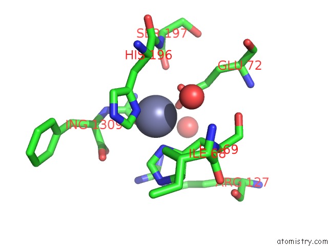
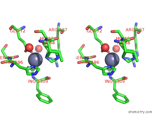
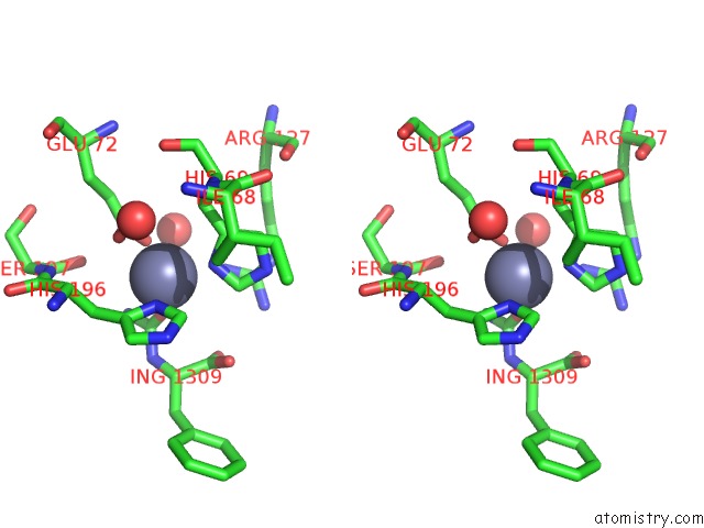
Zinc binding site 1 out of 4 in 1hdu
Go back to
Zinc binding site 1 out
of 4 in the Crystal Structure of Bovine Pancreatic Carboxypeptidase A Complexed with Aminocarbonylphenylalanine at 1.75 A
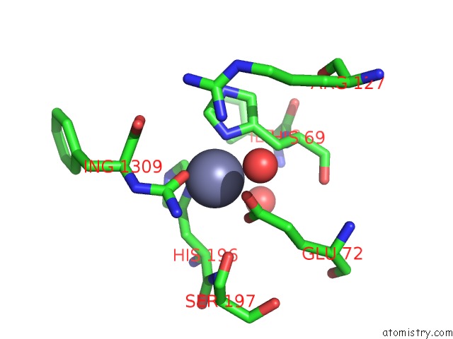
Mono view
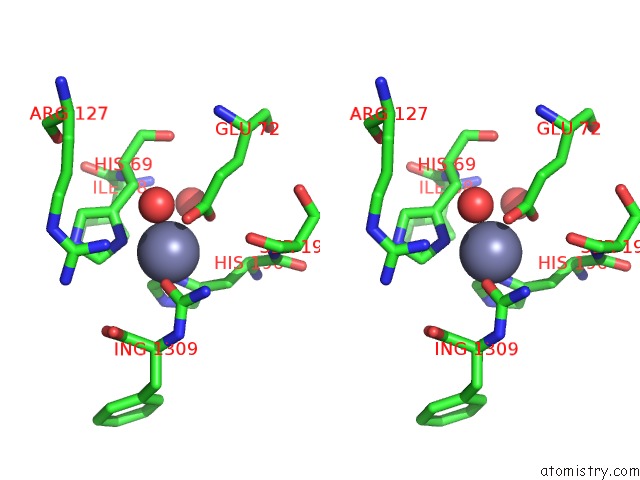
Stereo pair view
Mono view
Stereo pair view
A full contact list of Zinc with other atoms in the Zn binding
site number 1 of Crystal Structure of Bovine Pancreatic Carboxypeptidase A Complexed with Aminocarbonylphenylalanine at 1.75 A within 5.0Å range:
|
Zinc binding site 2 out of 4 in 1hdu
Go back to
Zinc binding site 2 out
of 4 in the Crystal Structure of Bovine Pancreatic Carboxypeptidase A Complexed with Aminocarbonylphenylalanine at 1.75 A
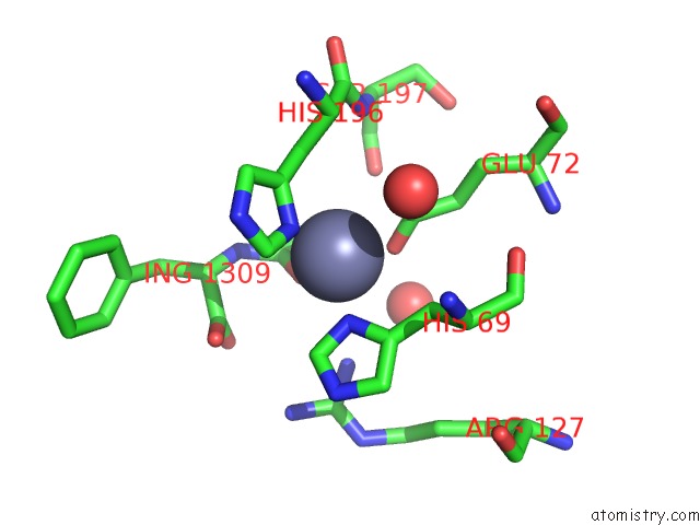
Mono view
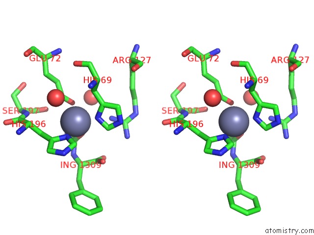
Stereo pair view
Mono view
Stereo pair view
A full contact list of Zinc with other atoms in the Zn binding
site number 2 of Crystal Structure of Bovine Pancreatic Carboxypeptidase A Complexed with Aminocarbonylphenylalanine at 1.75 A within 5.0Å range:
|
Zinc binding site 3 out of 4 in 1hdu
Go back to
Zinc binding site 3 out
of 4 in the Crystal Structure of Bovine Pancreatic Carboxypeptidase A Complexed with Aminocarbonylphenylalanine at 1.75 A
Mono view
Stereo pair view
Mono view
Stereo pair view
A full contact list of Zinc with other atoms in the Zn binding
site number 3 of Crystal Structure of Bovine Pancreatic Carboxypeptidase A Complexed with Aminocarbonylphenylalanine at 1.75 A within 5.0Å range:
|
Zinc binding site 4 out of 4 in 1hdu
Go back to
Zinc binding site 4 out
of 4 in the Crystal Structure of Bovine Pancreatic Carboxypeptidase A Complexed with Aminocarbonylphenylalanine at 1.75 A

Mono view
Stereo pair view
Mono view
Stereo pair view
A full contact list of Zinc with other atoms in the Zn binding
site number 4 of Crystal Structure of Bovine Pancreatic Carboxypeptidase A Complexed with Aminocarbonylphenylalanine at 1.75 A within 5.0Å range:
|
Reference:
J.H.Cho,
D.H.Kim,
S.J.Chung,
N.-C.Ha,
B.-H.Oh,
K.Y.Choi.
Insight Into the Stereochemistry in the Inhibition of Carboxypeptidase A with N- (Hydroxyaminocarbonyl)Phenylalanine: Binding Modes of An Enantiomeric Pair of the Inhibitor to Carboxypeptidase A Bioorg.Med.Chem. V. 10 2015 2002.
ISSN: ISSN 0968-0896
PubMed: 11937361
DOI: 10.1016/S0968-0896(01)00429-1
Page generated: Sun Oct 13 02:09:02 2024
ISSN: ISSN 0968-0896
PubMed: 11937361
DOI: 10.1016/S0968-0896(01)00429-1
Last articles
Zn in 9MJ5Zn in 9HNW
Zn in 9G0L
Zn in 9FNE
Zn in 9DZN
Zn in 9E0I
Zn in 9D32
Zn in 9DAK
Zn in 8ZXC
Zn in 8ZUF