Zinc »
PDB 1h71-1hld »
1h8l »
Zinc in PDB 1h8l: Duck Carboxypeptidase D Domain II in Complex with Gemsa
Protein crystallography data
The structure of Duck Carboxypeptidase D Domain II in Complex with Gemsa, PDB code: 1h8l
was solved by
F.X.Gomis-Rueth,
M.Coll,
F.X.Aviles,
J.Vendrell,
L.D.Fricker,
with X-Ray Crystallography technique. A brief refinement statistics is given in the table below:
| Resolution Low / High (Å) | 43.00 / 2.60 |
| Space group | P 21 3 |
| Cell size a, b, c (Å), α, β, γ (°) | 136.480, 136.480, 136.480, 90.00, 90.00, 90.00 |
| R / Rfree (%) | 19.9 / 23.9 |
Zinc Binding Sites:
The binding sites of Zinc atom in the Duck Carboxypeptidase D Domain II in Complex with Gemsa
(pdb code 1h8l). This binding sites where shown within
5.0 Angstroms radius around Zinc atom.
In total only one binding site of Zinc was determined in the Duck Carboxypeptidase D Domain II in Complex with Gemsa, PDB code: 1h8l:
In total only one binding site of Zinc was determined in the Duck Carboxypeptidase D Domain II in Complex with Gemsa, PDB code: 1h8l:
Zinc binding site 1 out of 1 in 1h8l
Go back to
Zinc binding site 1 out
of 1 in the Duck Carboxypeptidase D Domain II in Complex with Gemsa
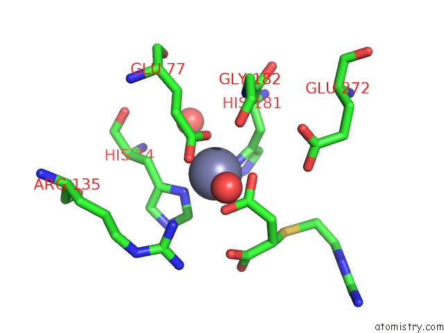
Mono view
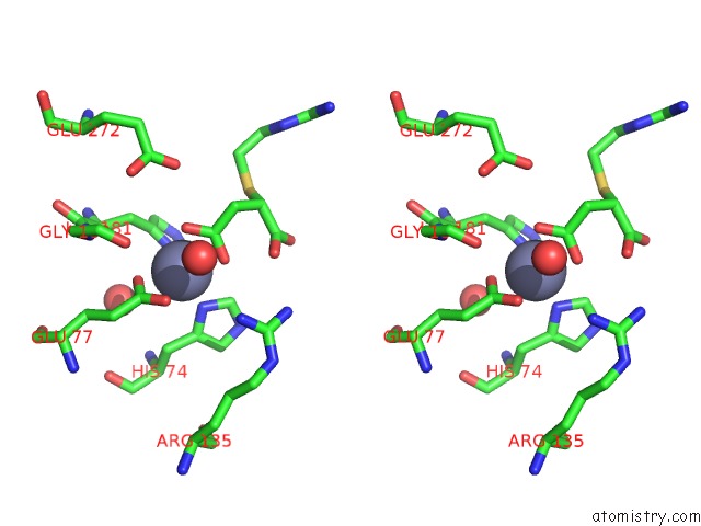
Stereo pair view
Mono view
Stereo pair view
A full contact list of Zinc with other atoms in the Zn binding
site number 1 of Duck Carboxypeptidase D Domain II in Complex with Gemsa within 5.0Å range:
|
Reference:
P.Aloy,
V.Companys,
J.Vendrell,
F.X.Aviles,
L.D.Fricker,
M.Coll,
F.X.Gomis-Ruth.
The Crystal Structure of the Inhibitor-Complexed Carboxypeptidase D Domain II and the Modeling of Regulatory Carboxypeptidases. J. Biol. Chem. V. 276 16177 2001.
ISSN: ISSN 0021-9258
PubMed: 11278909
DOI: 10.1074/JBC.M011457200
Page generated: Sun Oct 13 02:04:32 2024
ISSN: ISSN 0021-9258
PubMed: 11278909
DOI: 10.1074/JBC.M011457200
Last articles
Al in 3CF1Al in 3C7K
Al in 3B9R
Al in 3AB3
Al in 3BH7
Al in 3AR8
Al in 2YNM
Al in 2ZJY
Al in 2ZBG
Al in 2ZBD