Zinc »
PDB 1g45-1gkq »
1gjp »
Zinc in PDB 1gjp: Schiff-Base Complex of Yeast 5-Aminolaevulinic Acid Dehydratase with 4-Oxosebacic Acid
Enzymatic activity of Schiff-Base Complex of Yeast 5-Aminolaevulinic Acid Dehydratase with 4-Oxosebacic Acid
All present enzymatic activity of Schiff-Base Complex of Yeast 5-Aminolaevulinic Acid Dehydratase with 4-Oxosebacic Acid:
4.2.1.24;
4.2.1.24;
Protein crystallography data
The structure of Schiff-Base Complex of Yeast 5-Aminolaevulinic Acid Dehydratase with 4-Oxosebacic Acid, PDB code: 1gjp
was solved by
P.T.Erskine,
L.Coates,
R.Newbold,
A.A.Brindley,
S.P.Wood,
M.J.Warren,
J.B.Cooper,
P.M.Shoolingin-Jordan,
R.Neier,
with X-Ray Crystallography technique. A brief refinement statistics is given in the table below:
| Resolution Low / High (Å) | 30.40 / 1.80 |
| Space group | I 4 2 2 |
| Cell size a, b, c (Å), α, β, γ (°) | 103.500, 103.500, 168.200, 90.00, 90.00, 90.00 |
| R / Rfree (%) | 26.5 / 32.9 |
Zinc Binding Sites:
The binding sites of Zinc atom in the Schiff-Base Complex of Yeast 5-Aminolaevulinic Acid Dehydratase with 4-Oxosebacic Acid
(pdb code 1gjp). This binding sites where shown within
5.0 Angstroms radius around Zinc atom.
In total only one binding site of Zinc was determined in the Schiff-Base Complex of Yeast 5-Aminolaevulinic Acid Dehydratase with 4-Oxosebacic Acid, PDB code: 1gjp:
In total only one binding site of Zinc was determined in the Schiff-Base Complex of Yeast 5-Aminolaevulinic Acid Dehydratase with 4-Oxosebacic Acid, PDB code: 1gjp:
Zinc binding site 1 out of 1 in 1gjp
Go back to
Zinc binding site 1 out
of 1 in the Schiff-Base Complex of Yeast 5-Aminolaevulinic Acid Dehydratase with 4-Oxosebacic Acid
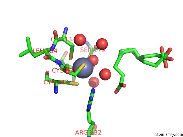
Mono view
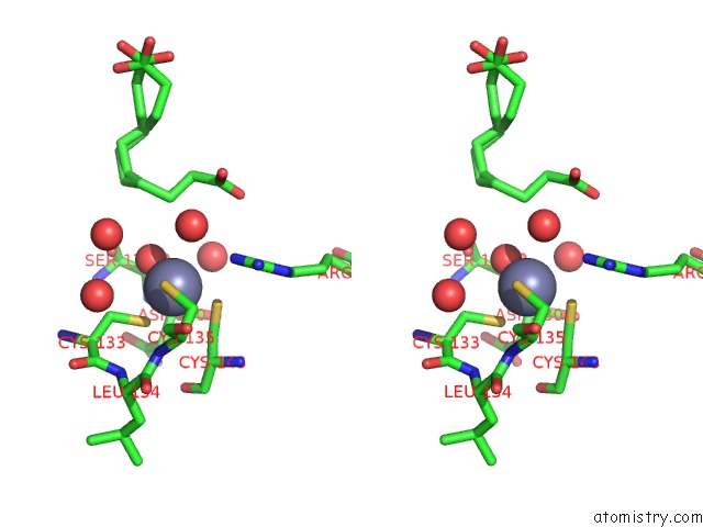
Stereo pair view
Mono view
Stereo pair view
A full contact list of Zinc with other atoms in the Zn binding
site number 1 of Schiff-Base Complex of Yeast 5-Aminolaevulinic Acid Dehydratase with 4-Oxosebacic Acid within 5.0Å range:
|
Reference:
P.T.Erskine,
L.Coates,
R.Newbold,
A.A.Brindley,
F.Stauffer,
S.P.Wood,
M.J.Warren,
J.B.Cooper,
P.M.Shoolingin-Jordan,
R.Neier.
The X-Ray Structure of Yeast 5-Aminolaevulinic Acid Dehydratase Complexed with Two Diacid Inhibitors Febs Lett. V. 503 196 2001.
ISSN: ISSN 0014-5793
PubMed: 11513881
DOI: 10.1016/S0014-5793(01)02721-1
Page generated: Sun Oct 13 01:32:40 2024
ISSN: ISSN 0014-5793
PubMed: 11513881
DOI: 10.1016/S0014-5793(01)02721-1
Last articles
As in 1C82As in 1BHL
As in 1D0C
As in 1BEH
As in 1B9D
As in 1B92
As in 1B9F
Ar in 7Q38
Ar in 6QAR
Ar in 6R1Q