Zinc »
PDB 1f6u-1fp0 »
1fjw »
Zinc in PDB 1fjw: Thermolysin (50 Mm Phenol Soaked)
Enzymatic activity of Thermolysin (50 Mm Phenol Soaked)
All present enzymatic activity of Thermolysin (50 Mm Phenol Soaked):
3.4.24.27;
3.4.24.27;
Protein crystallography data
The structure of Thermolysin (50 Mm Phenol Soaked), PDB code: 1fjw
was solved by
A.C.English,
C.R.Groom,
R.E.Hubbard,
with X-Ray Crystallography technique. A brief refinement statistics is given in the table below:
| Resolution Low / High (Å) | 15.00 / 1.90 |
| Space group | P 61 2 2 |
| Cell size a, b, c (Å), α, β, γ (°) | 93.960, 93.960, 130.915, 90.00, 90.00, 120.00 |
| R / Rfree (%) | 16.3 / 19.4 |
Other elements in 1fjw:
The structure of Thermolysin (50 Mm Phenol Soaked) also contains other interesting chemical elements:
| Calcium | (Ca) | 4 atoms |
Zinc Binding Sites:
The binding sites of Zinc atom in the Thermolysin (50 Mm Phenol Soaked)
(pdb code 1fjw). This binding sites where shown within
5.0 Angstroms radius around Zinc atom.
In total only one binding site of Zinc was determined in the Thermolysin (50 Mm Phenol Soaked), PDB code: 1fjw:
In total only one binding site of Zinc was determined in the Thermolysin (50 Mm Phenol Soaked), PDB code: 1fjw:
Zinc binding site 1 out of 1 in 1fjw
Go back to
Zinc binding site 1 out
of 1 in the Thermolysin (50 Mm Phenol Soaked)
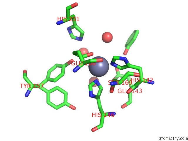
Mono view
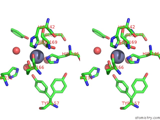
Stereo pair view
Mono view
Stereo pair view
A full contact list of Zinc with other atoms in the Zn binding
site number 1 of Thermolysin (50 Mm Phenol Soaked) within 5.0Å range:
|
Reference:
A.C.English,
C.R.Groom,
R.E.Hubbard.
Experimental and Computational Mapping of the Binding Surface of A Crystalline Protein. Protein Eng. V. 14 47 2001.
ISSN: ISSN 0269-2139
PubMed: 11287678
DOI: 10.1093/PROTEIN/14.1.47
Page generated: Sun Oct 13 01:00:18 2024
ISSN: ISSN 0269-2139
PubMed: 11287678
DOI: 10.1093/PROTEIN/14.1.47
Last articles
Al in 7NICAl in 7NIQ
Al in 7L07
Al in 7N77
Al in 7N73
Al in 7N72
Al in 7LVR
Al in 7KYB
Al in 7JL3
Al in 7KRO