Zinc »
PDB 1f6u-1fp0 »
1fbx »
Zinc in PDB 1fbx: Crystal Structure of Zinc-Containing E.Coli Gtp Cyclohydrolase I
Enzymatic activity of Crystal Structure of Zinc-Containing E.Coli Gtp Cyclohydrolase I
All present enzymatic activity of Crystal Structure of Zinc-Containing E.Coli Gtp Cyclohydrolase I:
3.5.4.16;
3.5.4.16;
Protein crystallography data
The structure of Crystal Structure of Zinc-Containing E.Coli Gtp Cyclohydrolase I, PDB code: 1fbx
was solved by
G.Auerbach,
A.Herrmann,
A.Bracher,
A.Bader,
M.Gutlich,
M.Fischer,
M.Neukamm,
H.Nar,
M.Garrido-Franco,
J.Richardson,
R.Huber,
A.Bacher,
with X-Ray Crystallography technique. A brief refinement statistics is given in the table below:
| Resolution Low / High (Å) | 14.98 / 2.80 |
| Space group | C 2 2 21 1 |
| Cell size a, b, c (Å), α, β, γ (°) | 227.690, 314.190, 132.570, 90.00, 90.00, 90.00 |
| R / Rfree (%) | 20.2 / 25.1 |
Other elements in 1fbx:
The structure of Crystal Structure of Zinc-Containing E.Coli Gtp Cyclohydrolase I also contains other interesting chemical elements:
| Chlorine | (Cl) | 15 atoms |
Zinc Binding Sites:
Pages:
>>> Page 1 <<< Page 2, Binding sites: 11 - 15;Binding sites:
The binding sites of Zinc atom in the Crystal Structure of Zinc-Containing E.Coli Gtp Cyclohydrolase I (pdb code 1fbx). This binding sites where shown within 5.0 Angstroms radius around Zinc atom.In total 15 binding sites of Zinc where determined in the Crystal Structure of Zinc-Containing E.Coli Gtp Cyclohydrolase I, PDB code: 1fbx:
Jump to Zinc binding site number: 1; 2; 3; 4; 5; 6; 7; 8; 9; 10;
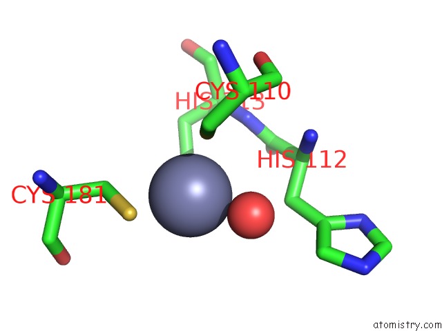
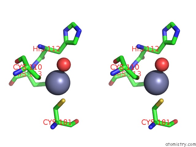
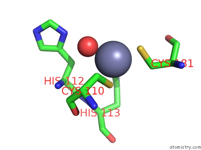
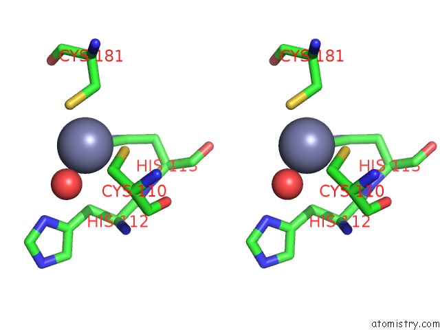
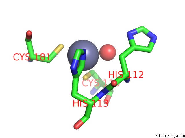
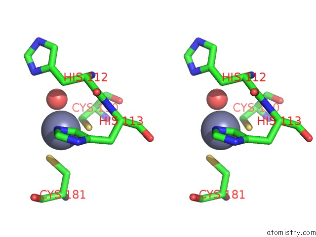
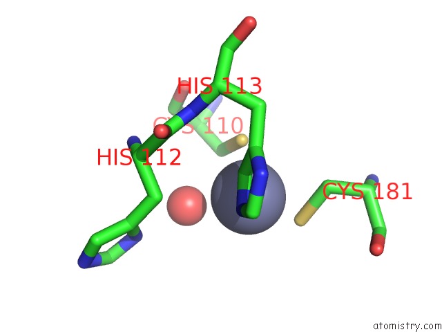
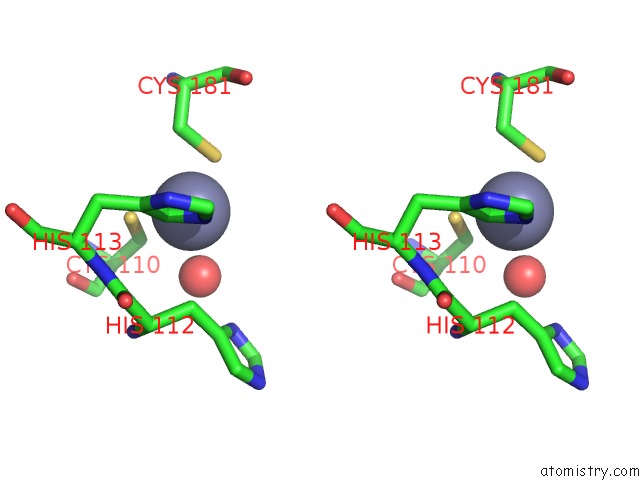
Zinc binding site 1 out of 15 in 1fbx
Go back to
Zinc binding site 1 out
of 15 in the Crystal Structure of Zinc-Containing E.Coli Gtp Cyclohydrolase I
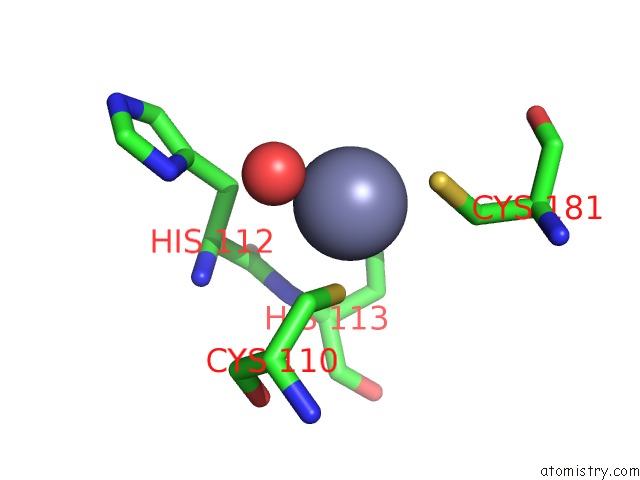
Mono view
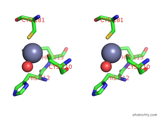
Stereo pair view
Mono view
Stereo pair view
A full contact list of Zinc with other atoms in the Zn binding
site number 1 of Crystal Structure of Zinc-Containing E.Coli Gtp Cyclohydrolase I within 5.0Å range:
|
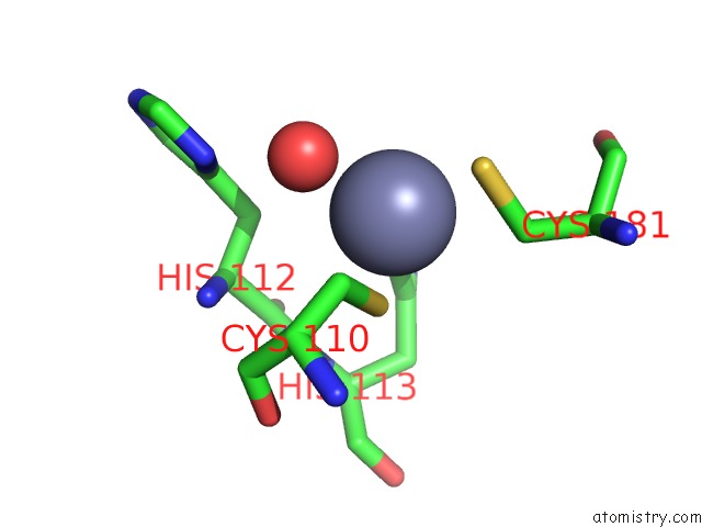
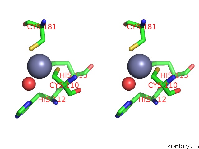
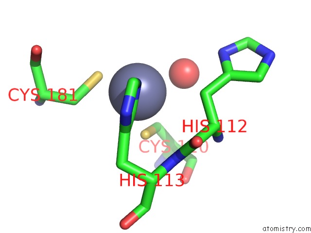
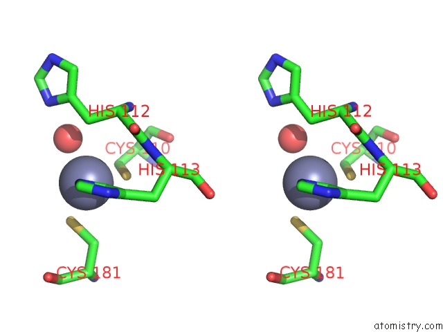
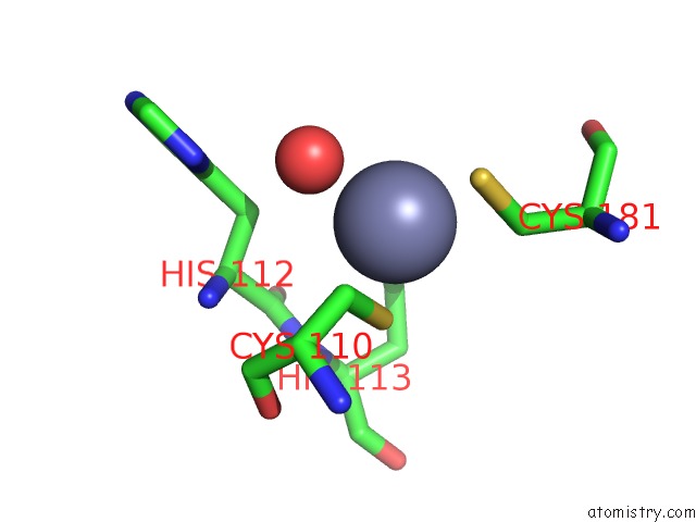
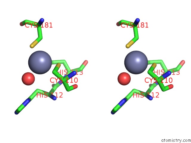
Zinc binding site 2 out of 15 in 1fbx
Go back to
Zinc binding site 2 out
of 15 in the Crystal Structure of Zinc-Containing E.Coli Gtp Cyclohydrolase I
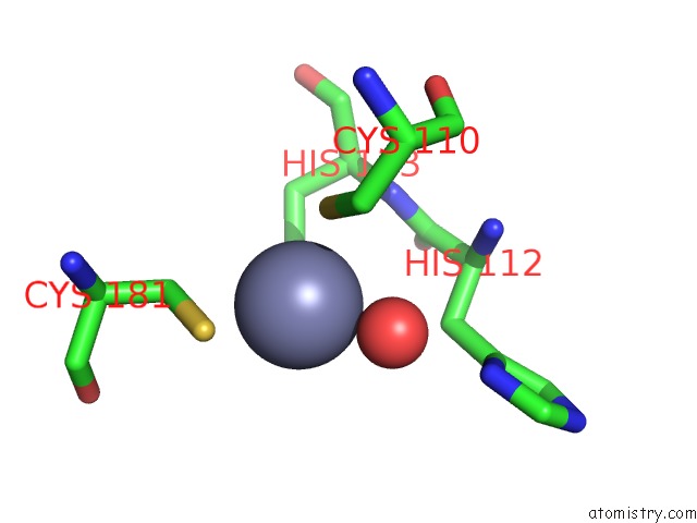
Mono view
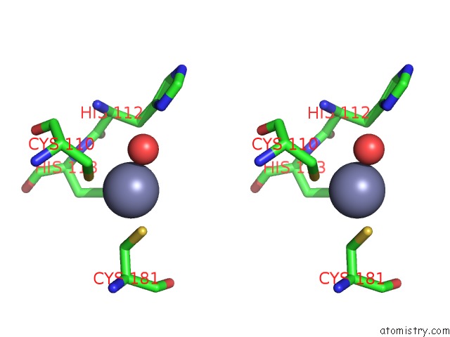
Stereo pair view
Mono view
Stereo pair view
A full contact list of Zinc with other atoms in the Zn binding
site number 2 of Crystal Structure of Zinc-Containing E.Coli Gtp Cyclohydrolase I within 5.0Å range:
|
Zinc binding site 3 out of 15 in 1fbx
Go back to
Zinc binding site 3 out
of 15 in the Crystal Structure of Zinc-Containing E.Coli Gtp Cyclohydrolase I
Mono view
Stereo pair view
Mono view
Stereo pair view
A full contact list of Zinc with other atoms in the Zn binding
site number 3 of Crystal Structure of Zinc-Containing E.Coli Gtp Cyclohydrolase I within 5.0Å range:
|
Zinc binding site 4 out of 15 in 1fbx
Go back to
Zinc binding site 4 out
of 15 in the Crystal Structure of Zinc-Containing E.Coli Gtp Cyclohydrolase I

Mono view

Stereo pair view
Mono view
Stereo pair view
A full contact list of Zinc with other atoms in the Zn binding
site number 4 of Crystal Structure of Zinc-Containing E.Coli Gtp Cyclohydrolase I within 5.0Å range:
|
Zinc binding site 5 out of 15 in 1fbx
Go back to
Zinc binding site 5 out
of 15 in the Crystal Structure of Zinc-Containing E.Coli Gtp Cyclohydrolase I
Mono view
Stereo pair view
Mono view
Stereo pair view
A full contact list of Zinc with other atoms in the Zn binding
site number 5 of Crystal Structure of Zinc-Containing E.Coli Gtp Cyclohydrolase I within 5.0Å range:
|
Zinc binding site 6 out of 15 in 1fbx
Go back to
Zinc binding site 6 out
of 15 in the Crystal Structure of Zinc-Containing E.Coli Gtp Cyclohydrolase I
Mono view
Stereo pair view
Mono view
Stereo pair view
A full contact list of Zinc with other atoms in the Zn binding
site number 6 of Crystal Structure of Zinc-Containing E.Coli Gtp Cyclohydrolase I within 5.0Å range:
|
Zinc binding site 7 out of 15 in 1fbx
Go back to
Zinc binding site 7 out
of 15 in the Crystal Structure of Zinc-Containing E.Coli Gtp Cyclohydrolase I
Mono view
Stereo pair view
Mono view
Stereo pair view
A full contact list of Zinc with other atoms in the Zn binding
site number 7 of Crystal Structure of Zinc-Containing E.Coli Gtp Cyclohydrolase I within 5.0Å range:
|
Zinc binding site 8 out of 15 in 1fbx
Go back to
Zinc binding site 8 out
of 15 in the Crystal Structure of Zinc-Containing E.Coli Gtp Cyclohydrolase I
Mono view
Stereo pair view
Mono view
Stereo pair view
A full contact list of Zinc with other atoms in the Zn binding
site number 8 of Crystal Structure of Zinc-Containing E.Coli Gtp Cyclohydrolase I within 5.0Å range:
|
Zinc binding site 9 out of 15 in 1fbx
Go back to
Zinc binding site 9 out
of 15 in the Crystal Structure of Zinc-Containing E.Coli Gtp Cyclohydrolase I
Mono view
Stereo pair view
Mono view
Stereo pair view
A full contact list of Zinc with other atoms in the Zn binding
site number 9 of Crystal Structure of Zinc-Containing E.Coli Gtp Cyclohydrolase I within 5.0Å range:
|
Zinc binding site 10 out of 15 in 1fbx
Go back to
Zinc binding site 10 out
of 15 in the Crystal Structure of Zinc-Containing E.Coli Gtp Cyclohydrolase I
Mono view
Stereo pair view
Mono view
Stereo pair view
A full contact list of Zinc with other atoms in the Zn binding
site number 10 of Crystal Structure of Zinc-Containing E.Coli Gtp Cyclohydrolase I within 5.0Å range:
|
Reference:
G.Auerbach,
A.Herrmann,
A.Bracher,
G.Bader,
M.Gutlich,
M.Fischer,
M.Neukamm,
M.Garrido-Franco,
J.Richardson,
H.Nar,
R.Huber,
A.Bacher.
Zinc Plays A Key Role in Human and Bacterial Gtp Cyclohydrolase I. Proc.Natl.Acad.Sci.Usa V. 97 13567 2000.
ISSN: ISSN 0027-8424
PubMed: 11087827
DOI: 10.1073/PNAS.240463497
Page generated: Sun Oct 13 00:55:22 2024
ISSN: ISSN 0027-8424
PubMed: 11087827
DOI: 10.1073/PNAS.240463497
Last articles
Zn in 9MJ5Zn in 9HNW
Zn in 9G0L
Zn in 9FNE
Zn in 9DZN
Zn in 9E0I
Zn in 9D32
Zn in 9DAK
Zn in 8ZXC
Zn in 8ZUF