Zinc »
PDB 1ed8-1evl »
1eou »
Zinc in PDB 1eou: Crystal Structure of Human Carbonic Anhydrase II Complexed with An Anticonvulsant Sugar Sulfamate
Enzymatic activity of Crystal Structure of Human Carbonic Anhydrase II Complexed with An Anticonvulsant Sugar Sulfamate
All present enzymatic activity of Crystal Structure of Human Carbonic Anhydrase II Complexed with An Anticonvulsant Sugar Sulfamate:
4.2.1.1;
4.2.1.1;
Protein crystallography data
The structure of Crystal Structure of Human Carbonic Anhydrase II Complexed with An Anticonvulsant Sugar Sulfamate, PDB code: 1eou
was solved by
R.Recacha,
M.J.Costanzo,
B.E.Maryanoff,
D.Chattopadhyay,
with X-Ray Crystallography technique. A brief refinement statistics is given in the table below:
| Resolution Low / High (Å) | 14.99 / 2.10 |
| Space group | P 1 21 1 |
| Cell size a, b, c (Å), α, β, γ (°) | 42.275, 41.390, 72.509, 90.00, 104.11, 90.00 |
| R / Rfree (%) | 17.6 / 22.9 |
Zinc Binding Sites:
The binding sites of Zinc atom in the Crystal Structure of Human Carbonic Anhydrase II Complexed with An Anticonvulsant Sugar Sulfamate
(pdb code 1eou). This binding sites where shown within
5.0 Angstroms radius around Zinc atom.
In total only one binding site of Zinc was determined in the Crystal Structure of Human Carbonic Anhydrase II Complexed with An Anticonvulsant Sugar Sulfamate, PDB code: 1eou:
In total only one binding site of Zinc was determined in the Crystal Structure of Human Carbonic Anhydrase II Complexed with An Anticonvulsant Sugar Sulfamate, PDB code: 1eou:
Zinc binding site 1 out of 1 in 1eou
Go back to
Zinc binding site 1 out
of 1 in the Crystal Structure of Human Carbonic Anhydrase II Complexed with An Anticonvulsant Sugar Sulfamate
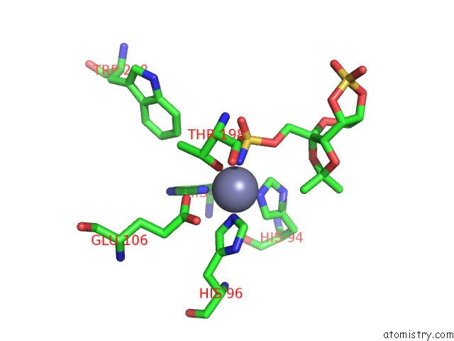
Mono view
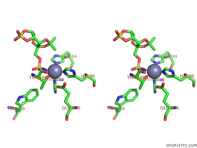
Stereo pair view
Mono view
Stereo pair view
A full contact list of Zinc with other atoms in the Zn binding
site number 1 of Crystal Structure of Human Carbonic Anhydrase II Complexed with An Anticonvulsant Sugar Sulfamate within 5.0Å range:
|
Reference:
R.Recacha,
M.J.Costanzo,
B.E.Maryanoff,
D.Chattopadhyay.
Crystal Structure of Human Carbonic Anhydrase II Complexed with An Anti-Convulsant Sugar Sulphamate. Biochem.J. V. 361 437 2002.
ISSN: ISSN 0264-6021
PubMed: 11802772
DOI: 10.1042/0264-6021:3610437
Page generated: Sun Oct 13 00:21:47 2024
ISSN: ISSN 0264-6021
PubMed: 11802772
DOI: 10.1042/0264-6021:3610437
Last articles
Zn in 9MJ5Zn in 9HNW
Zn in 9G0L
Zn in 9FNE
Zn in 9DZN
Zn in 9E0I
Zn in 9D32
Zn in 9DAK
Zn in 8ZXC
Zn in 8ZUF