Zinc »
PDB 1e0e-1ed6 »
1ed5 »
Zinc in PDB 1ed5: Bovine Endothelial Nitric Oxide Synthase Heme Domain Complexed with Nna(H4B Free)
Enzymatic activity of Bovine Endothelial Nitric Oxide Synthase Heme Domain Complexed with Nna(H4B Free)
All present enzymatic activity of Bovine Endothelial Nitric Oxide Synthase Heme Domain Complexed with Nna(H4B Free):
1.14.13.39;
1.14.13.39;
Protein crystallography data
The structure of Bovine Endothelial Nitric Oxide Synthase Heme Domain Complexed with Nna(H4B Free), PDB code: 1ed5
was solved by
C.S.Raman,
H.Li,
P.Martasek,
G.J.Southan,
B.S.S.Masters,
T.L.Poulos,
with X-Ray Crystallography technique. A brief refinement statistics is given in the table below:
| Resolution Low / High (Å) | 39.46 / 1.80 |
| Space group | P 21 21 21 |
| Cell size a, b, c (Å), α, β, γ (°) | 58.826, 106.417, 156.163, 90.00, 90.00, 90.00 |
| R / Rfree (%) | 20.6 / 23.8 |
Other elements in 1ed5:
The structure of Bovine Endothelial Nitric Oxide Synthase Heme Domain Complexed with Nna(H4B Free) also contains other interesting chemical elements:
| Arsenic | (As) | 2 atoms |
| Iron | (Fe) | 2 atoms |
Zinc Binding Sites:
The binding sites of Zinc atom in the Bovine Endothelial Nitric Oxide Synthase Heme Domain Complexed with Nna(H4B Free)
(pdb code 1ed5). This binding sites where shown within
5.0 Angstroms radius around Zinc atom.
In total only one binding site of Zinc was determined in the Bovine Endothelial Nitric Oxide Synthase Heme Domain Complexed with Nna(H4B Free), PDB code: 1ed5:
In total only one binding site of Zinc was determined in the Bovine Endothelial Nitric Oxide Synthase Heme Domain Complexed with Nna(H4B Free), PDB code: 1ed5:
Zinc binding site 1 out of 1 in 1ed5
Go back to
Zinc binding site 1 out
of 1 in the Bovine Endothelial Nitric Oxide Synthase Heme Domain Complexed with Nna(H4B Free)
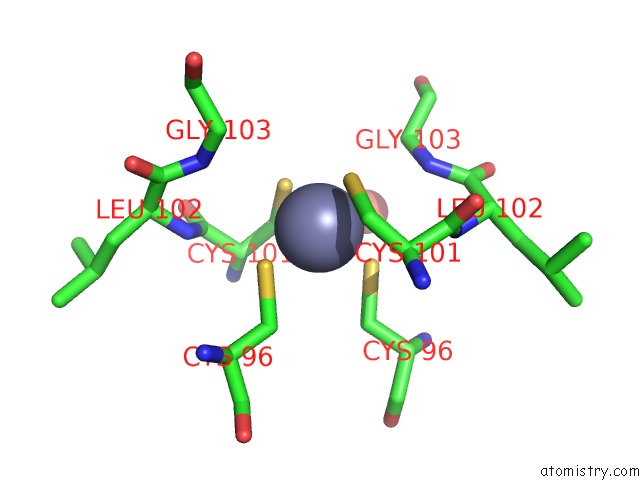
Mono view
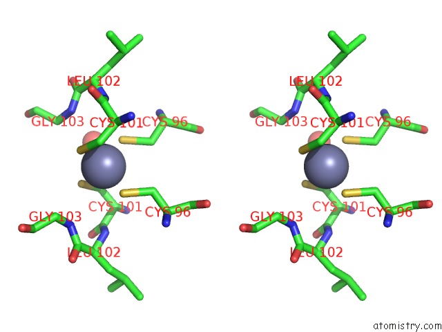
Stereo pair view
Mono view
Stereo pair view
A full contact list of Zinc with other atoms in the Zn binding
site number 1 of Bovine Endothelial Nitric Oxide Synthase Heme Domain Complexed with Nna(H4B Free) within 5.0Å range:
|
Reference:
C.S.Raman,
H.Li,
P.Martasek,
G.Southan,
B.S.Masters,
T.L.Poulos.
Crystal Structure of Nitric Oxide Synthase Bound to Nitro Indazole Reveals A Novel Inactivation Mechanism. Biochemistry V. 40 13448 2001.
ISSN: ISSN 0006-2960
PubMed: 11695891
DOI: 10.1021/BI010957U
Page generated: Sun Oct 13 00:08:17 2024
ISSN: ISSN 0006-2960
PubMed: 11695891
DOI: 10.1021/BI010957U
Last articles
Zn in 9MJ5Zn in 9HNW
Zn in 9G0L
Zn in 9FNE
Zn in 9DZN
Zn in 9E0I
Zn in 9D32
Zn in 9DAK
Zn in 8ZXC
Zn in 8ZUF