Zinc »
PDB 1e0e-1ed6 »
1ebo »
Zinc in PDB 1ebo: Crystal Structure of the Ebola Virus Membrane-Fusion Subunit, GP2, From the Envelope Glycoprotein Ectodomain
Protein crystallography data
The structure of Crystal Structure of the Ebola Virus Membrane-Fusion Subunit, GP2, From the Envelope Glycoprotein Ectodomain, PDB code: 1ebo
was solved by
W.Weissenhorn,
A.Carfi,
K.H.Lee,
J.J.Skehel,
D.C.Wiley,
with X-Ray Crystallography technique. A brief refinement statistics is given in the table below:
| Resolution Low / High (Å) | 20.00 / 3.00 |
| Space group | C 1 2 1 |
| Cell size a, b, c (Å), α, β, γ (°) | 171.702, 32.691, 168.863, 90.00, 119.23, 90.00 |
| R / Rfree (%) | 23.9 / 25.6 |
Other elements in 1ebo:
The structure of Crystal Structure of the Ebola Virus Membrane-Fusion Subunit, GP2, From the Envelope Glycoprotein Ectodomain also contains other interesting chemical elements:
| Chlorine | (Cl) | 2 atoms |
Zinc Binding Sites:
The binding sites of Zinc atom in the Crystal Structure of the Ebola Virus Membrane-Fusion Subunit, GP2, From the Envelope Glycoprotein Ectodomain
(pdb code 1ebo). This binding sites where shown within
5.0 Angstroms radius around Zinc atom.
In total 3 binding sites of Zinc where determined in the Crystal Structure of the Ebola Virus Membrane-Fusion Subunit, GP2, From the Envelope Glycoprotein Ectodomain, PDB code: 1ebo:
Jump to Zinc binding site number: 1; 2; 3;
In total 3 binding sites of Zinc where determined in the Crystal Structure of the Ebola Virus Membrane-Fusion Subunit, GP2, From the Envelope Glycoprotein Ectodomain, PDB code: 1ebo:
Jump to Zinc binding site number: 1; 2; 3;
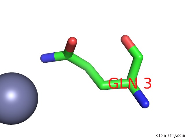
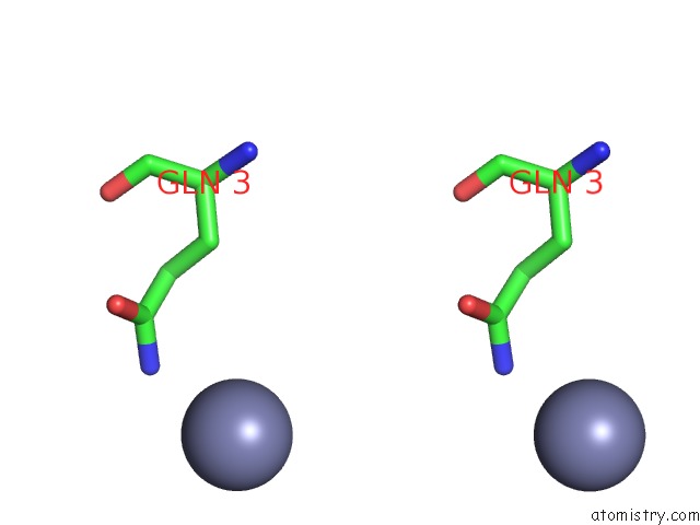
Zinc binding site 1 out of 3 in 1ebo
Go back to
Zinc binding site 1 out
of 3 in the Crystal Structure of the Ebola Virus Membrane-Fusion Subunit, GP2, From the Envelope Glycoprotein Ectodomain
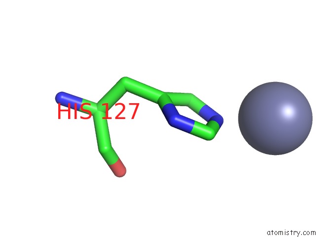
Mono view
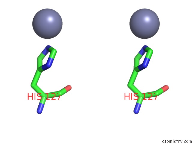
Stereo pair view
Mono view
Stereo pair view
A full contact list of Zinc with other atoms in the Zn binding
site number 1 of Crystal Structure of the Ebola Virus Membrane-Fusion Subunit, GP2, From the Envelope Glycoprotein Ectodomain within 5.0Å range:
|
Zinc binding site 2 out of 3 in 1ebo
Go back to
Zinc binding site 2 out
of 3 in the Crystal Structure of the Ebola Virus Membrane-Fusion Subunit, GP2, From the Envelope Glycoprotein Ectodomain
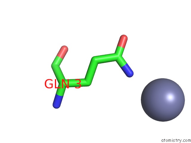
Mono view
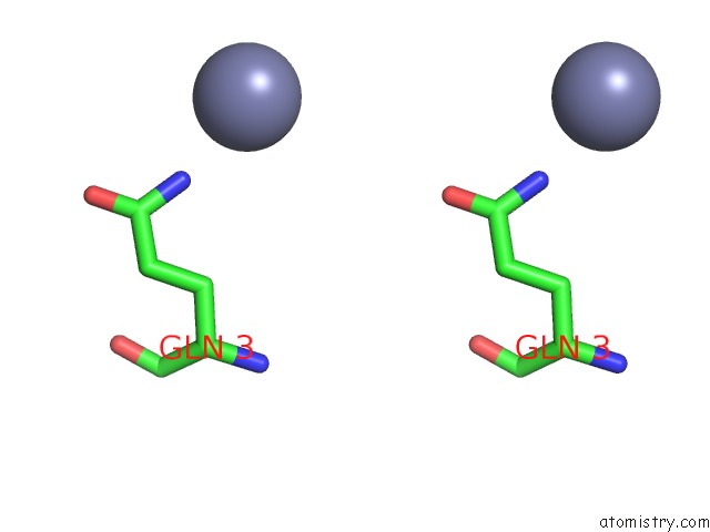
Stereo pair view
Mono view
Stereo pair view
A full contact list of Zinc with other atoms in the Zn binding
site number 2 of Crystal Structure of the Ebola Virus Membrane-Fusion Subunit, GP2, From the Envelope Glycoprotein Ectodomain within 5.0Å range:
|
Zinc binding site 3 out of 3 in 1ebo
Go back to
Zinc binding site 3 out
of 3 in the Crystal Structure of the Ebola Virus Membrane-Fusion Subunit, GP2, From the Envelope Glycoprotein Ectodomain
Mono view
Stereo pair view
Mono view
Stereo pair view
A full contact list of Zinc with other atoms in the Zn binding
site number 3 of Crystal Structure of the Ebola Virus Membrane-Fusion Subunit, GP2, From the Envelope Glycoprotein Ectodomain within 5.0Å range:
|
Reference:
W.Weissenhorn,
A.Carfi,
K.H.Lee,
J.J.Skehel,
D.C.Wiley.
Crystal Structure of the Ebola Virus Membrane Fusion Subunit, GP2, From the Envelope Glycoprotein Ectodomain. Mol.Cell V. 2 605 1998.
ISSN: ISSN 1097-2765
PubMed: 9844633
DOI: 10.1016/S1097-2765(00)80159-8
Page generated: Sun Oct 13 00:07:06 2024
ISSN: ISSN 1097-2765
PubMed: 9844633
DOI: 10.1016/S1097-2765(00)80159-8
Last articles
Zn in 9MJ5Zn in 9HNW
Zn in 9G0L
Zn in 9FNE
Zn in 9DZN
Zn in 9E0I
Zn in 9D32
Zn in 9DAK
Zn in 8ZXC
Zn in 8ZUF