Zinc »
PDB 1e0e-1ed6 »
1e4m »
Zinc in PDB 1e4m: Myrosinase From Sinapis Alba
Enzymatic activity of Myrosinase From Sinapis Alba
All present enzymatic activity of Myrosinase From Sinapis Alba:
3.2.1.147;
3.2.1.147;
Protein crystallography data
The structure of Myrosinase From Sinapis Alba, PDB code: 1e4m
was solved by
W.P.Burmeister,
with X-Ray Crystallography technique. A brief refinement statistics is given in the table below:
| Resolution Low / High (Å) | 10.00 / 1.20 |
| Space group | C 2 2 21 |
| Cell size a, b, c (Å), α, β, γ (°) | 135.300, 137.200, 80.600, 90.00, 90.00, 90.00 |
| R / Rfree (%) | 12.4 / 14.2 |
Zinc Binding Sites:
The binding sites of Zinc atom in the Myrosinase From Sinapis Alba
(pdb code 1e4m). This binding sites where shown within
5.0 Angstroms radius around Zinc atom.
In total only one binding site of Zinc was determined in the Myrosinase From Sinapis Alba, PDB code: 1e4m:
In total only one binding site of Zinc was determined in the Myrosinase From Sinapis Alba, PDB code: 1e4m:
Zinc binding site 1 out of 1 in 1e4m
Go back to
Zinc binding site 1 out
of 1 in the Myrosinase From Sinapis Alba
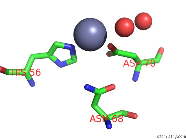
Mono view
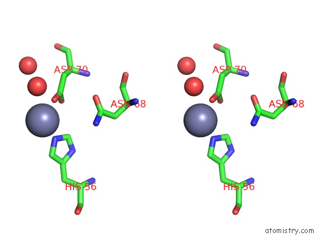
Stereo pair view
Mono view
Stereo pair view
A full contact list of Zinc with other atoms in the Zn binding
site number 1 of Myrosinase From Sinapis Alba within 5.0Å range:
|
Reference:
W.P.Burmeister,
S.Cottaz,
P.Rollin,
A.Vasella,
B.Henrissat.
High Resolution X-Ray Crystallography Shows That Ascorbate Is A Cofactor For Myrosinase and Substitutes For the Function of the Catalytic Base J.Biol.Chem. V. 275 39385 2000.
ISSN: ISSN 0021-9258
PubMed: 10978344
DOI: 10.1074/JBC.M006796200
Page generated: Sun Oct 13 00:02:41 2024
ISSN: ISSN 0021-9258
PubMed: 10978344
DOI: 10.1074/JBC.M006796200
Last articles
Zn in 9MJ5Zn in 9HNW
Zn in 9G0L
Zn in 9FNE
Zn in 9DZN
Zn in 9E0I
Zn in 9D32
Zn in 9DAK
Zn in 8ZXC
Zn in 8ZUF