Zinc »
PDB 1do5-1e08 »
1dqs »
Zinc in PDB 1dqs: Crystal Structure of Dehydroquinate Synthase (Dhqs) Complexed with Carbaphosphonate, Nad+ and ZN2+
Enzymatic activity of Crystal Structure of Dehydroquinate Synthase (Dhqs) Complexed with Carbaphosphonate, Nad+ and ZN2+
All present enzymatic activity of Crystal Structure of Dehydroquinate Synthase (Dhqs) Complexed with Carbaphosphonate, Nad+ and ZN2+:
4.6.1.3;
4.6.1.3;
Protein crystallography data
The structure of Crystal Structure of Dehydroquinate Synthase (Dhqs) Complexed with Carbaphosphonate, Nad+ and ZN2+, PDB code: 1dqs
was solved by
E.P.Carpenter,
A.R.Hawkins,
J.W.Frost,
K.A.Brown,
with X-Ray Crystallography technique. A brief refinement statistics is given in the table below:
| Resolution Low / High (Å) | 25.00 / 1.80 |
| Space group | P 21 21 21 |
| Cell size a, b, c (Å), α, β, γ (°) | 67.677, 80.810, 143.513, 90.00, 90.00, 90.00 |
| R / Rfree (%) | 17.4 / 22.4 |
Other elements in 1dqs:
The structure of Crystal Structure of Dehydroquinate Synthase (Dhqs) Complexed with Carbaphosphonate, Nad+ and ZN2+ also contains other interesting chemical elements:
| Chlorine | (Cl) | 1 atom |
Zinc Binding Sites:
The binding sites of Zinc atom in the Crystal Structure of Dehydroquinate Synthase (Dhqs) Complexed with Carbaphosphonate, Nad+ and ZN2+
(pdb code 1dqs). This binding sites where shown within
5.0 Angstroms radius around Zinc atom.
In total 2 binding sites of Zinc where determined in the Crystal Structure of Dehydroquinate Synthase (Dhqs) Complexed with Carbaphosphonate, Nad+ and ZN2+, PDB code: 1dqs:
Jump to Zinc binding site number: 1; 2;
In total 2 binding sites of Zinc where determined in the Crystal Structure of Dehydroquinate Synthase (Dhqs) Complexed with Carbaphosphonate, Nad+ and ZN2+, PDB code: 1dqs:
Jump to Zinc binding site number: 1; 2;
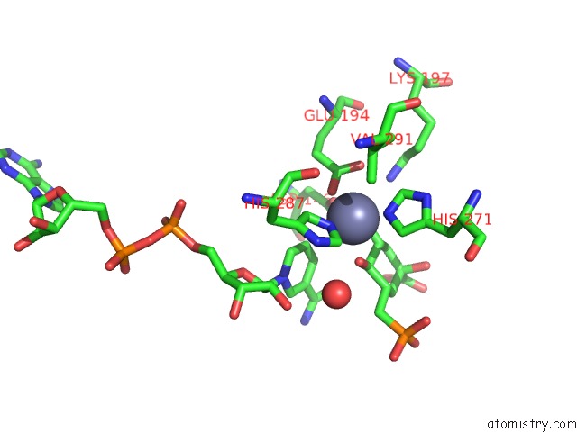
Zinc binding site 1 out of 2 in 1dqs
Go back to
Zinc binding site 1 out
of 2 in the Crystal Structure of Dehydroquinate Synthase (Dhqs) Complexed with Carbaphosphonate, Nad+ and ZN2+
Mono view

Stereo pair view
Mono view
Stereo pair view
A full contact list of Zinc with other atoms in the Zn binding
site number 1 of Crystal Structure of Dehydroquinate Synthase (Dhqs) Complexed with Carbaphosphonate, Nad+ and ZN2+ within 5.0Å range:
|
Zinc binding site 2 out of 2 in 1dqs
Go back to
Zinc binding site 2 out
of 2 in the Crystal Structure of Dehydroquinate Synthase (Dhqs) Complexed with Carbaphosphonate, Nad+ and ZN2+

Mono view

Stereo pair view
Mono view
Stereo pair view
A full contact list of Zinc with other atoms in the Zn binding
site number 2 of Crystal Structure of Dehydroquinate Synthase (Dhqs) Complexed with Carbaphosphonate, Nad+ and ZN2+ within 5.0Å range:
|
Reference:
E.P.Carpenter,
A.R.Hawkins,
J.W.Frost,
K.A.Brown.
Structure of Dehydroquinate Synthase Reveals An Active Site Capable of Multistep Catalysis. Nature V. 394 299 1998.
ISSN: ISSN 0028-0836
PubMed: 9685163
DOI: 10.1038/28431
Page generated: Sat Oct 12 23:48:51 2024
ISSN: ISSN 0028-0836
PubMed: 9685163
DOI: 10.1038/28431
Last articles
Zn in 9MJ5Zn in 9HNW
Zn in 9G0L
Zn in 9FNE
Zn in 9DZN
Zn in 9E0I
Zn in 9D32
Zn in 9DAK
Zn in 8ZXC
Zn in 8ZUF