Zinc »
PDB 1cp6-1d4u »
1d1w »
Zinc in PDB 1d1w: Bovine Endothelial Nitric Oxide Synthase Heme Domain Complexed with 2- Aminothiazoline (H4B Bound)
Enzymatic activity of Bovine Endothelial Nitric Oxide Synthase Heme Domain Complexed with 2- Aminothiazoline (H4B Bound)
All present enzymatic activity of Bovine Endothelial Nitric Oxide Synthase Heme Domain Complexed with 2- Aminothiazoline (H4B Bound):
1.14.13.39;
1.14.13.39;
Protein crystallography data
The structure of Bovine Endothelial Nitric Oxide Synthase Heme Domain Complexed with 2- Aminothiazoline (H4B Bound), PDB code: 1d1w
was solved by
H.Li,
C.S.Raman,
P.Martasek,
V.Kral,
B.S.S.Masters,
T.L.Poulos,
with X-Ray Crystallography technique. A brief refinement statistics is given in the table below:
| Resolution Low / High (Å) | 50.00 / 2.00 |
| Space group | P 21 21 21 |
| Cell size a, b, c (Å), α, β, γ (°) | 58.460, 106.320, 156.330, 90.00, 90.00, 90.00 |
| R / Rfree (%) | 20.4 / 25 |
Other elements in 1d1w:
The structure of Bovine Endothelial Nitric Oxide Synthase Heme Domain Complexed with 2- Aminothiazoline (H4B Bound) also contains other interesting chemical elements:
| Arsenic | (As) | 2 atoms |
| Iron | (Fe) | 2 atoms |
Zinc Binding Sites:
The binding sites of Zinc atom in the Bovine Endothelial Nitric Oxide Synthase Heme Domain Complexed with 2- Aminothiazoline (H4B Bound)
(pdb code 1d1w). This binding sites where shown within
5.0 Angstroms radius around Zinc atom.
In total only one binding site of Zinc was determined in the Bovine Endothelial Nitric Oxide Synthase Heme Domain Complexed with 2- Aminothiazoline (H4B Bound), PDB code: 1d1w:
In total only one binding site of Zinc was determined in the Bovine Endothelial Nitric Oxide Synthase Heme Domain Complexed with 2- Aminothiazoline (H4B Bound), PDB code: 1d1w:
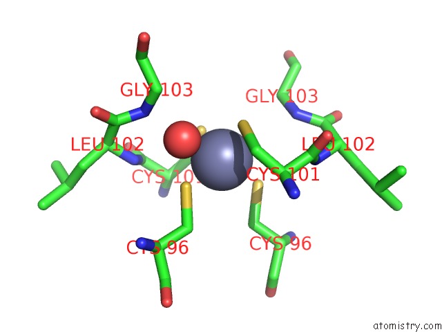
Zinc binding site 1 out of 1 in 1d1w
Go back to
Zinc binding site 1 out
of 1 in the Bovine Endothelial Nitric Oxide Synthase Heme Domain Complexed with 2- Aminothiazoline (H4B Bound)
Mono view

Stereo pair view
Mono view
Stereo pair view
A full contact list of Zinc with other atoms in the Zn binding
site number 1 of Bovine Endothelial Nitric Oxide Synthase Heme Domain Complexed with 2- Aminothiazoline (H4B Bound) within 5.0Å range:
|
Reference:
H.Li,
C.S.Raman,
P.Martasek,
V.Kral,
B.S.Masters,
T.L.Poulos.
Mapping the Active Site Polarity in Structures of Endothelial Nitric Oxide Synthase Heme Domain Complexed with Isothioureas. J.Inorg.Biochem. V. 81 133 2000.
ISSN: ISSN 0162-0134
PubMed: 11051558
DOI: 10.1016/S0162-0134(00)00099-4
Page generated: Sat Oct 12 23:27:00 2024
ISSN: ISSN 0162-0134
PubMed: 11051558
DOI: 10.1016/S0162-0134(00)00099-4
Last articles
Al in 8R1AAl in 8Q75
Al in 8OIE
Al in 8OX6
Al in 8OX5
Al in 8OP8
Al in 8OP6
Al in 8OP5
Al in 8OOS
Al in 8OO9