Zinc »
PDB 1bj6-1bv3 »
1bsk »
Zinc in PDB 1bsk: Zinc Deformylase Inhibitor Complex From E.Coli
Enzymatic activity of Zinc Deformylase Inhibitor Complex From E.Coli
All present enzymatic activity of Zinc Deformylase Inhibitor Complex From E.Coli:
3.5.1.27;
3.5.1.27;
Protein crystallography data
The structure of Zinc Deformylase Inhibitor Complex From E.Coli, PDB code: 1bsk
was solved by
B.Hao,
W.Gong,
P.T.Rajagopalan,
Y.Hu,
D.Pei,
M.K.Chan,
with X-Ray Crystallography technique. A brief refinement statistics is given in the table below:
| Resolution Low / High (Å) | 20.00 / 3.00 |
| Space group | P 65 2 2 |
| Cell size a, b, c (Å), α, β, γ (°) | 99.170, 99.170, 110.755, 90.00, 90.00, 120.00 |
| R / Rfree (%) | 16.7 / 22.1 |
Zinc Binding Sites:
The binding sites of Zinc atom in the Zinc Deformylase Inhibitor Complex From E.Coli
(pdb code 1bsk). This binding sites where shown within
5.0 Angstroms radius around Zinc atom.
In total only one binding site of Zinc was determined in the Zinc Deformylase Inhibitor Complex From E.Coli, PDB code: 1bsk:
In total only one binding site of Zinc was determined in the Zinc Deformylase Inhibitor Complex From E.Coli, PDB code: 1bsk:
Zinc binding site 1 out of 1 in 1bsk
Go back to
Zinc binding site 1 out
of 1 in the Zinc Deformylase Inhibitor Complex From E.Coli
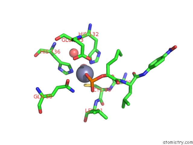
Mono view
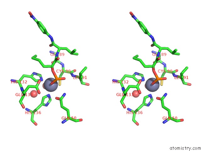
Stereo pair view
Mono view
Stereo pair view
A full contact list of Zinc with other atoms in the Zn binding
site number 1 of Zinc Deformylase Inhibitor Complex From E.Coli within 5.0Å range:
|
Reference:
B.Hao,
W.Gong,
P.T.Rajagopalan,
Y.Zhou,
D.Pei,
M.K.Chan.
Structural Basis For the Design of Antibiotics Targeting Peptide Deformylase. Biochemistry V. 38 4712 1999.
ISSN: ISSN 0006-2960
PubMed: 10200158
DOI: 10.1021/BI982594C
Page generated: Sat Oct 12 22:43:17 2024
ISSN: ISSN 0006-2960
PubMed: 10200158
DOI: 10.1021/BI982594C
Last articles
As in 1JZWAs in 1ITG
As in 1II9
As in 1HYZ
As in 1II0
As in 1IHU
As in 1I83
As in 1I9T
As in 1I9S
As in 1HYV