Zinc »
PDB 1adf-1axg »
1ak0 »
Zinc in PDB 1ak0: P1 Nuclease in Complex with A Substrate Analog
Enzymatic activity of P1 Nuclease in Complex with A Substrate Analog
All present enzymatic activity of P1 Nuclease in Complex with A Substrate Analog:
3.1.30.1;
3.1.30.1;
Protein crystallography data
The structure of P1 Nuclease in Complex with A Substrate Analog, PDB code: 1ak0
was solved by
C.Romier,
D.Suck,
with X-Ray Crystallography technique. A brief refinement statistics is given in the table below:
| Resolution Low / High (Å) | 8.00 / 1.80 |
| Space group | P 21 21 21 |
| Cell size a, b, c (Å), α, β, γ (°) | 41.980, 74.040, 102.130, 90.00, 90.00, 90.00 |
| R / Rfree (%) | 20.7 / 23.5 |
Zinc Binding Sites:
The binding sites of Zinc atom in the P1 Nuclease in Complex with A Substrate Analog
(pdb code 1ak0). This binding sites where shown within
5.0 Angstroms radius around Zinc atom.
In total 4 binding sites of Zinc where determined in the P1 Nuclease in Complex with A Substrate Analog, PDB code: 1ak0:
Jump to Zinc binding site number: 1; 2; 3; 4;
In total 4 binding sites of Zinc where determined in the P1 Nuclease in Complex with A Substrate Analog, PDB code: 1ak0:
Jump to Zinc binding site number: 1; 2; 3; 4;
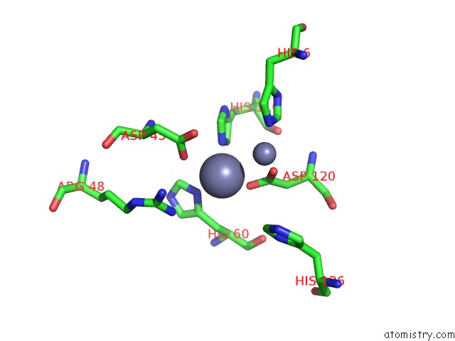
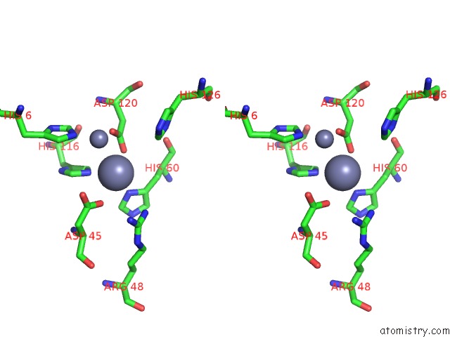
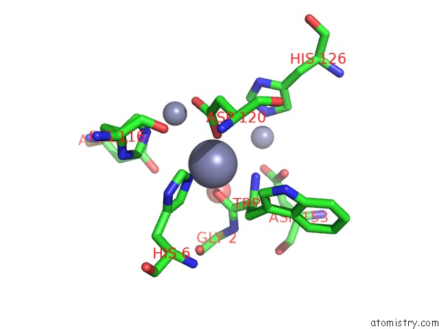
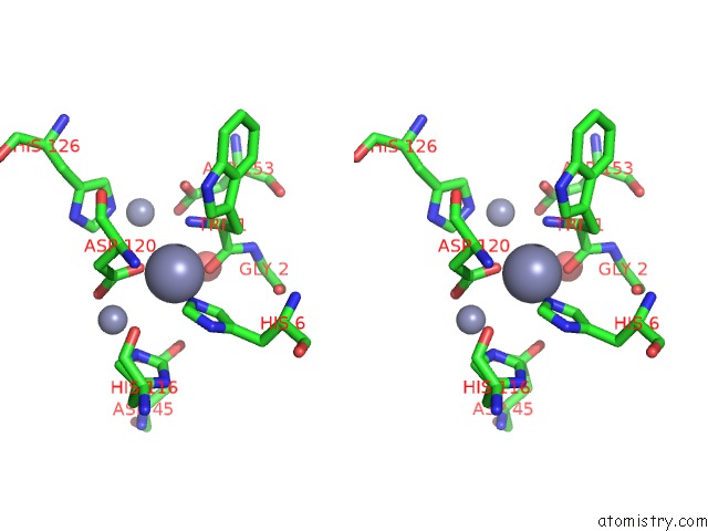
Zinc binding site 1 out of 4 in 1ak0
Go back to
Zinc binding site 1 out
of 4 in the P1 Nuclease in Complex with A Substrate Analog
Mono view
Stereo pair view
Mono view
Stereo pair view
A full contact list of Zinc with other atoms in the Zn binding
site number 1 of P1 Nuclease in Complex with A Substrate Analog within 5.0Å range:
|
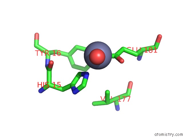
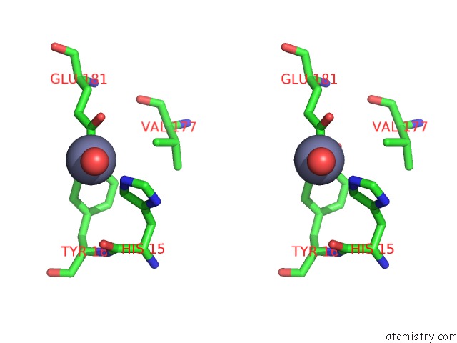
Zinc binding site 2 out of 4 in 1ak0
Go back to
Zinc binding site 2 out
of 4 in the P1 Nuclease in Complex with A Substrate Analog
Mono view
Stereo pair view
Mono view
Stereo pair view
A full contact list of Zinc with other atoms in the Zn binding
site number 2 of P1 Nuclease in Complex with A Substrate Analog within 5.0Å range:
|
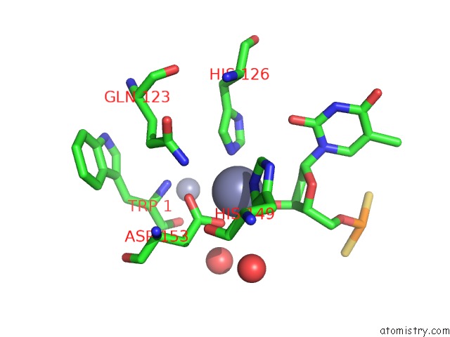
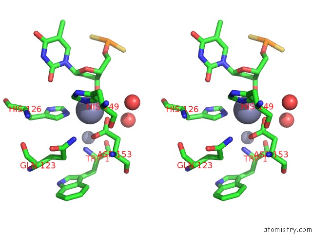
Zinc binding site 3 out of 4 in 1ak0
Go back to
Zinc binding site 3 out
of 4 in the P1 Nuclease in Complex with A Substrate Analog
Mono view
Stereo pair view
Mono view
Stereo pair view
A full contact list of Zinc with other atoms in the Zn binding
site number 3 of P1 Nuclease in Complex with A Substrate Analog within 5.0Å range:
|
Zinc binding site 4 out of 4 in 1ak0
Go back to
Zinc binding site 4 out
of 4 in the P1 Nuclease in Complex with A Substrate Analog
Mono view
Stereo pair view
Mono view
Stereo pair view
A full contact list of Zinc with other atoms in the Zn binding
site number 4 of P1 Nuclease in Complex with A Substrate Analog within 5.0Å range:
|
Reference:
C.Romier,
R.Dominguez,
A.Lahm,
O.Dahl,
D.Suck.
Recognition of Single-Stranded Dna By Nuclease P1: High Resolution Crystal Structures of Complexes with Substrate Analogs. Proteins V. 32 414 1998.
ISSN: ISSN 0887-3585
PubMed: 9726413
DOI: 10.1002/(SICI)1097-0134(19980901)32:4<414::AID-PROT2>3.3.CO;2-5
Page generated: Sat Oct 12 22:06:33 2024
ISSN: ISSN 0887-3585
PubMed: 9726413
DOI: 10.1002/(SICI)1097-0134(19980901)32:4<414::AID-PROT2>3.3.CO;2-5
Last articles
Zn in 9MJ5Zn in 9HNW
Zn in 9G0L
Zn in 9FNE
Zn in 9DZN
Zn in 9E0I
Zn in 9D32
Zn in 9DAK
Zn in 8ZXC
Zn in 8ZUF