Zinc »
PDB 1adf-1axg »
1adu »
Zinc in PDB 1adu: Early E2A Dna-Binding Protein
Protein crystallography data
The structure of Early E2A Dna-Binding Protein, PDB code: 1adu
was solved by
P.A.Tucker,
P.N.Kanellopoulos,
D.Tsernoglou,
P.C.Van Der Vliet,
with X-Ray Crystallography technique. A brief refinement statistics is given in the table below:
| Resolution Low / High (Å) | 6.00 / 3.00 |
| Space group | P 21 21 21 |
| Cell size a, b, c (Å), α, β, γ (°) | 61.000, 91.200, 149.400, 90.00, 90.00, 90.00 |
| R / Rfree (%) | 20.1 / 29.6 |
Zinc Binding Sites:
The binding sites of Zinc atom in the Early E2A Dna-Binding Protein
(pdb code 1adu). This binding sites where shown within
5.0 Angstroms radius around Zinc atom.
In total 4 binding sites of Zinc where determined in the Early E2A Dna-Binding Protein, PDB code: 1adu:
Jump to Zinc binding site number: 1; 2; 3; 4;
In total 4 binding sites of Zinc where determined in the Early E2A Dna-Binding Protein, PDB code: 1adu:
Jump to Zinc binding site number: 1; 2; 3; 4;
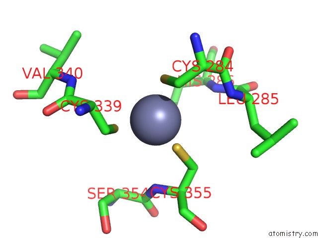
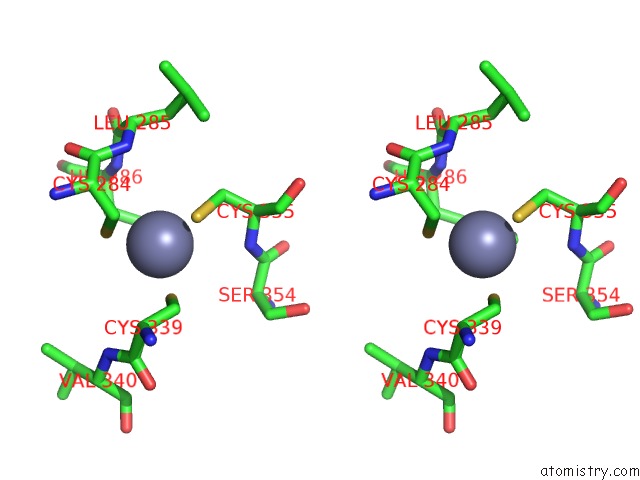
Zinc binding site 1 out of 4 in 1adu
Go back to
Zinc binding site 1 out
of 4 in the Early E2A Dna-Binding Protein
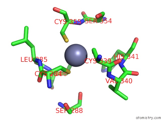
Mono view
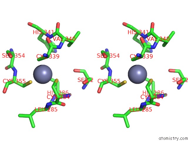
Stereo pair view
Mono view
Stereo pair view
A full contact list of Zinc with other atoms in the Zn binding
site number 1 of Early E2A Dna-Binding Protein within 5.0Å range:
|
Zinc binding site 2 out of 4 in 1adu
Go back to
Zinc binding site 2 out
of 4 in the Early E2A Dna-Binding Protein
Mono view
Stereo pair view
Mono view
Stereo pair view
A full contact list of Zinc with other atoms in the Zn binding
site number 2 of Early E2A Dna-Binding Protein within 5.0Å range:
|
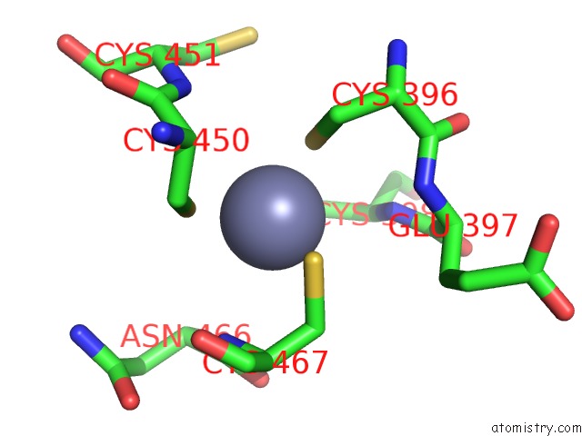
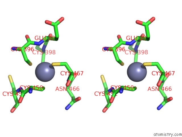
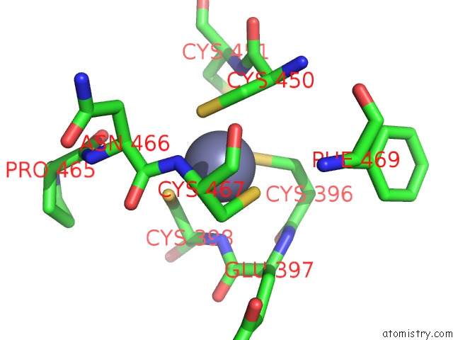
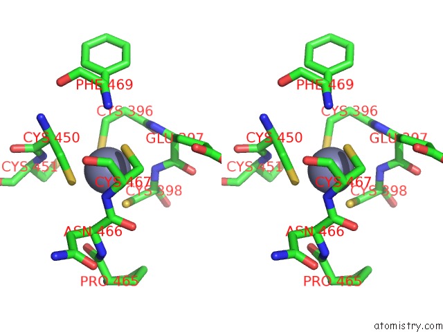
Zinc binding site 3 out of 4 in 1adu
Go back to
Zinc binding site 3 out
of 4 in the Early E2A Dna-Binding Protein
Mono view
Stereo pair view
Mono view
Stereo pair view
A full contact list of Zinc with other atoms in the Zn binding
site number 3 of Early E2A Dna-Binding Protein within 5.0Å range:
|
Zinc binding site 4 out of 4 in 1adu
Go back to
Zinc binding site 4 out
of 4 in the Early E2A Dna-Binding Protein
Mono view
Stereo pair view
Mono view
Stereo pair view
A full contact list of Zinc with other atoms in the Zn binding
site number 4 of Early E2A Dna-Binding Protein within 5.0Å range:
|
Reference:
P.N.Kanellopoulos,
D.Tsernoglou,
P.C.Van Der Vliet,
P.A.Tucker.
Alternative Arrangements of the Protein Chain Are Possible For the Adenovirus Single-Stranded Dna Binding Protein. J.Mol.Biol. V. 257 1 1996.
ISSN: ISSN 0022-2836
PubMed: 8632448
DOI: 10.1006/JMBI.1996.0141
Page generated: Sat Oct 12 21:59:14 2024
ISSN: ISSN 0022-2836
PubMed: 8632448
DOI: 10.1006/JMBI.1996.0141
Last articles
Zn in 9MJ5Zn in 9HNW
Zn in 9G0L
Zn in 9FNE
Zn in 9DZN
Zn in 9E0I
Zn in 9D32
Zn in 9DAK
Zn in 8ZXC
Zn in 8ZUF