Zinc »
PDB 12ca-1add »
1a4l »
Zinc in PDB 1a4l: Ada Structure Complexed with Deoxycoformycin at pH 7.0
Enzymatic activity of Ada Structure Complexed with Deoxycoformycin at pH 7.0
All present enzymatic activity of Ada Structure Complexed with Deoxycoformycin at pH 7.0:
3.5.4.4;
3.5.4.4;
Protein crystallography data
The structure of Ada Structure Complexed with Deoxycoformycin at pH 7.0, PDB code: 1a4l
was solved by
Z.Wang,
F.A.Quiocho,
with X-Ray Crystallography technique. A brief refinement statistics is given in the table below:
| Resolution Low / High (Å) | 8.00 / 2.60 |
| Space group | P 1 21 1 |
| Cell size a, b, c (Å), α, β, γ (°) | 88.929, 93.355, 101.953, 90.00, 102.94, 90.00 |
| R / Rfree (%) | 18.5 / 27.2 |
Zinc Binding Sites:
The binding sites of Zinc atom in the Ada Structure Complexed with Deoxycoformycin at pH 7.0
(pdb code 1a4l). This binding sites where shown within
5.0 Angstroms radius around Zinc atom.
In total 4 binding sites of Zinc where determined in the Ada Structure Complexed with Deoxycoformycin at pH 7.0, PDB code: 1a4l:
Jump to Zinc binding site number: 1; 2; 3; 4;
In total 4 binding sites of Zinc where determined in the Ada Structure Complexed with Deoxycoformycin at pH 7.0, PDB code: 1a4l:
Jump to Zinc binding site number: 1; 2; 3; 4;
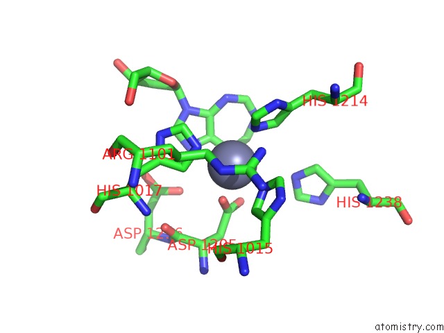
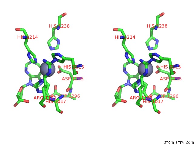
Zinc binding site 1 out of 4 in 1a4l
Go back to
Zinc binding site 1 out
of 4 in the Ada Structure Complexed with Deoxycoformycin at pH 7.0
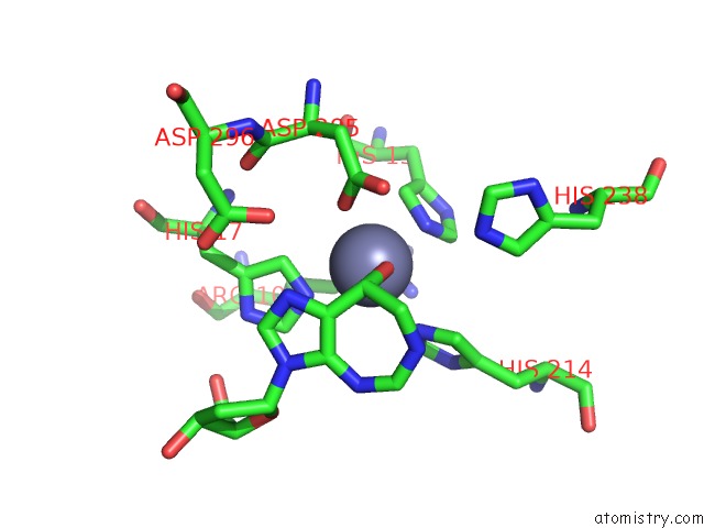
Mono view
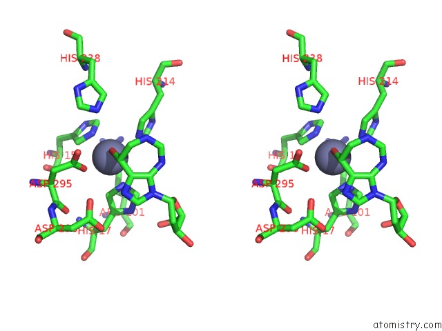
Stereo pair view
Mono view
Stereo pair view
A full contact list of Zinc with other atoms in the Zn binding
site number 1 of Ada Structure Complexed with Deoxycoformycin at pH 7.0 within 5.0Å range:
|
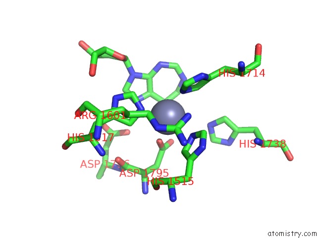
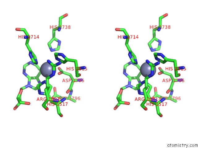
Zinc binding site 2 out of 4 in 1a4l
Go back to
Zinc binding site 2 out
of 4 in the Ada Structure Complexed with Deoxycoformycin at pH 7.0
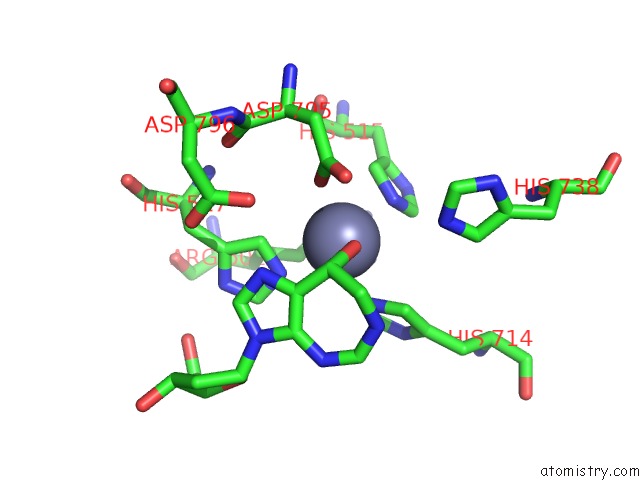
Mono view
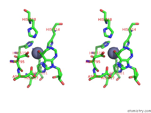
Stereo pair view
Mono view
Stereo pair view
A full contact list of Zinc with other atoms in the Zn binding
site number 2 of Ada Structure Complexed with Deoxycoformycin at pH 7.0 within 5.0Å range:
|
Zinc binding site 3 out of 4 in 1a4l
Go back to
Zinc binding site 3 out
of 4 in the Ada Structure Complexed with Deoxycoformycin at pH 7.0
Mono view
Stereo pair view
Mono view
Stereo pair view
A full contact list of Zinc with other atoms in the Zn binding
site number 3 of Ada Structure Complexed with Deoxycoformycin at pH 7.0 within 5.0Å range:
|
Zinc binding site 4 out of 4 in 1a4l
Go back to
Zinc binding site 4 out
of 4 in the Ada Structure Complexed with Deoxycoformycin at pH 7.0
Mono view
Stereo pair view
Mono view
Stereo pair view
A full contact list of Zinc with other atoms in the Zn binding
site number 4 of Ada Structure Complexed with Deoxycoformycin at pH 7.0 within 5.0Å range:
|
Reference:
Z.Wang,
F.A.Quiocho.
Complexes of Adenosine Deaminase with Two Potent Inhibitors: X-Ray Structures in Four Independent Molecules at pH of Maximum Activity. Biochemistry V. 37 8314 1998.
ISSN: ISSN 0006-2960
PubMed: 9622483
DOI: 10.1021/BI980324O
Page generated: Sat Oct 12 21:46:30 2024
ISSN: ISSN 0006-2960
PubMed: 9622483
DOI: 10.1021/BI980324O
Last articles
Zn in 9MJ5Zn in 9HNW
Zn in 9G0L
Zn in 9FNE
Zn in 9DZN
Zn in 9E0I
Zn in 9D32
Zn in 9DAK
Zn in 8ZXC
Zn in 8ZUF